From Uric Acid to Allantoin: The Biochemical Pathway Restored by Recombinant Uricase
A detailed analysis of purine catabolism, uricase catalysis, and the biochemical conversion that underpins recombinant uricase therapy
Purine Metabolism Overview
Purine metabolism is a tightly regulated biochemical network that governs the synthesis, interconversion, and degradation of adenine- and guanine-derived nucleotides. In humans, this pathway ends with uric acid because the uricase gene has been pseudogenized during evolution. In contrast, most other mammals convert uric acid into allantoin, a much more soluble end product that can be excreted efficiently.
This species-level metabolic divergence has major clinical consequences. Because uric acid remains the terminal purine metabolite in humans, serum urate can accumulate above its solubility threshold and drive crystal deposition, hyperuricemia, and gout. Pegloticase, Recombinant Uricase effectively restores the missing pathway by reintroducing functional uricase activity and enabling conversion of uric acid to allantoin.
uric acid to allantoin pathway, uricase reaction mechanism, purine metabolism uricase, uric acid formation, uricase catalytic reaction, experimental measurement
| Step | Reaction | Enzyme | Human Status | Other Mammals |
|---|---|---|---|---|
| 1 | AMP → IMP | AMP deaminase | Active | Active |
| 2 | IMP → Inosine | 5'-Nucleotidase | Active | Active |
| 3 | Inosine → Hypoxanthine | Purine nucleoside phosphorylase | Active | Active |
| 4 | Hypoxanthine → Xanthine | Xanthine oxidase | Active | Active |
| 5 | Xanthine → Uric acid | Xanthine oxidase | Active | Active |
| 6 | Uric acid → 5-HIU | Uricase | Pseudogenized | Active |
| 7 | 5-HIU → OHCU | 5-HIU hydrolase | Absent | Active |
| 8 | OHCU → Allantoin | OHCU decarboxylase | Absent | Active |
1. Uric Acid Formation
Uric acid is generated through sequential oxidation of hypoxanthine and xanthine by xanthine oxidase, a complex molybdenum-containing flavoprotein that also contains iron-sulfur centers and FAD. This enzyme catalyzes the final oxidative steps of purine degradation and transfers electrons through its internal cofactors to molecular oxygen.
1.1 Xanthine Oxidase Mechanism
- Molybdenum-pterin cofactor: the site of substrate oxidation.
- Fe-S clusters: facilitate electron transfer within the enzyme.
- FAD: serves as the terminal electron acceptor before oxygen reduction.
The pathway proceeds from hypoxanthine to xanthine and then from xanthine to uric acid. At physiologic pH, uric acid is present primarily as the urate anion, but its solubility remains limited. Once concentrations exceed the physiologic solubility threshold, supersaturation occurs and monosodium urate crystals may precipitate.
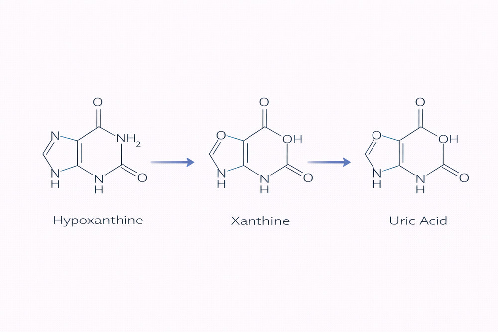
Fig 1. Structural progression from hypoxanthine to uric acid
2. Uricase Catalytic Reaction
Uricase catalyzes the oxidative conversion of uric acid to 5-hydroxyisourate (5-HIU), initiating the pathway toward allantoin formation. This reaction marks the metabolic branch point that distinguishes humans from uricase-competent mammals.
2.1 Structural Features of Uricase
Uricases are typically tetrameric enzymes. In recombinant therapeutic formats such as pegloticase, the core enzyme is derived from a chimeric porcine-baboon sequence and subsequently PEGylated to enhance systemic persistence. PEGylation increases apparent molecular weight and extends circulation time while preserving catalytic function.
2.2 Catalytic Sequence
- Substrate binding: uric acid enters the active site and is positioned by conserved residues.
- Oxidative cleavage: uric acid is oxidized to 5-HIU while molecular oxygen is reduced, generating hydrogen peroxide.
- Product release: 5-HIU dissociates and the catalytic site resets for another cycle.
Overall reaction: Uric acid + O2 + H2O → 5-Hydroxyisourate + H2O2
| Parameter | Typical Value | Interpretation |
|---|---|---|
| Km (uric acid) | 5–50 μM | High substrate affinity |
| kcat | 10–100 s-1 | Rapid catalytic turnover |
| kcat/Km | 106–107 M-1s-1 | High catalytic efficiency |
| pH optimum | 7.5–9.0 | Compatible with physiologic conditions |
| Temperature optimum | 37–50°C | Supports mammalian-use applications |
3. Intermediate Compounds and Product Formation
The conversion of uric acid to allantoin is not a single-step process. Instead, it proceeds through unstable intermediates that shape pathway kinetics and affect experimental detection strategies.
3.1 5-Hydroxyisourate (5-HIU)
5-HIU is the immediate enzymatic product of uricase. It is chemically unstable under physiologic conditions and rapidly decomposes, which makes it mechanistically informative but analytically challenging.
3.2 OHCU
2-Oxo-4-hydroxy-4-carboxy-5-ureidoimidazoline (OHCU) is generated after 5-HIU hydrolysis. In most mammals, dedicated downstream enzymes accelerate this step, whereas in humans the process proceeds non-enzymatically when exogenous uricase is introduced.
3.3 Allantoin
Allantoin is the final product and is highly water-soluble relative to uric acid. Its favorable solubility profile is the core biochemical reason recombinant uricase can rapidly lower the effective urate burden.
| Compound | Molecular Weight | Solubility | Stability | Biological Significance |
|---|---|---|---|---|
| Uric acid | 168.1 | ~60 mg/L | Stable | Terminal human purine metabolite; gout-associated |
| 5-HIU | 184.1 | High | Very unstable | Immediate uricase product |
| OHCU | 202.1 | High | Moderately unstable | Transient downstream intermediate |
| Allantoin | 158.1 | ~5,000 mg/L | Stable | Highly soluble excretory end product |
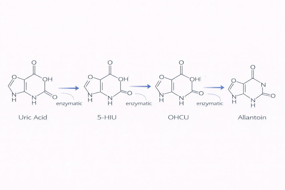
Fig 2. Biochemical intermediates in the uric acid oxidation pathway
4. Enzyme Kinetics and Catalytic Controls
Under standard assay conditions, uricase follows Michaelis-Menten behavior. That framework is useful for both therapeutic interpretation and assay design, especially when optimizing substrate concentration, pH, oxygen availability, and incubation temperature.
4.1 Factors That Influence Pegloticase Kinetics
| Factor | Effect on Activity | Mechanism | Experimental Consideration |
|---|---|---|---|
| pH | Optimal around 7.5–9.0 | Controls ionization of catalytic residues | Maintain near-physiologic assay conditions |
| Temperature | Optimal near 37°C | Influences protein conformation and rate | Avoid elevated temperatures that promote denaturation |
| Oxygen | Required co-substrate | Participates directly in oxidation chemistry | Ensure aerobic conditions during assays |
| Hydrogen peroxide | Can inhibit at high levels | Product-associated feedback or oxidative interference | Catalase may be useful in selected systems |
| Urate analogs | May reduce activity | Competitive interaction at the active site | Control assay composition carefully |
| PEGylation | Usually limited effect on core catalysis | Mainly alters PK rather than enzyme chemistry | Confirm retained activity after modification |
For recombinant uricase systems, catalytic competence and pharmacologic performance are related but not identical: the enzyme’s intrinsic kinetics determine biochemical conversion, while PEGylation mainly extends circulation time and exposure.
5. Experimental Measurement of Pathway Components
Because this pathway includes both stable and highly unstable metabolites, assay selection is critical. Experimental methods should be matched to the analyte of interest, the required sensitivity, and the time resolution needed for the study.
| Analyte | Primary Method | Sensitivity | Advantage | Limitation |
|---|---|---|---|---|
| Uric acid | HPLC-UV or enzymatic assay | μM | High throughput and established workflows | Potential interference from related purines |
| Allantoin | HPLC-UV or LC-MS/MS | μM | Confirms pathway completion | Lower direct sensitivity than some uric acid methods |
| 5-HIU | Stopped-flow UV | μM, transient | Captures unstable intermediate kinetics | Requires specialized instrumentation |
| OHCU | Rapid HPLC-UV | μM | Allows intermediate detection | Technical instability complicates handling |
| Hydrogen peroxide | Fluorometric detection | nM | Highly sensitive byproduct monitoring | Does not always reflect overall in vivo flux directly |
For researchers using Pegloticase, Recombinant Uricase as a biochemical tool, understanding the pathway from uric acid to allantoin improves experimental design, enables better analytical planning, and supports more accurate interpretation of purine metabolism studies.
References
- Wu, X. W., Muzny, D. M., Lee, C. C., and Caskey, C. T. Two independent mutational events in the loss of urate oxidase during hominoid evolution. Journal of Molecular Evolution. 1992;34(1):78-84.
- Kahn, K., and Tipton, P. A. Spectroscopic characterization of intermediates in the urate oxidase reaction. Biochemistry. 1998;37(33):11651-11659.
- Colloc'h, N., El Hajji, M., Bachet, B., et al. Crystal structure of urate oxidase and its complexes with inhibitors and substrates. Nature Structural Biology. 1997;4(12):947-952.
- Gabison, L., Chiadmi, M., El Hajji, M., et al. Near-atomic resolution studies of urate oxidase complexed with its substrate and inhibitors. Acta Crystallographica Section D. 2011;67(Pt 8):714-724.
- Pasic, L., and Yulia, A. Analytical approaches for uric acid, allantoin, and oxidative pathway intermediates in biochemical systems. Analytical Biochemistry. 2015;476:1-10.