Uricase Activity Assay Protocol
A standardized workflow for measuring pegloticase activity through uric acid degradation
Abstract
The quantitative assessment of uricase activity is essential for quality control, research applications, and therapeutic monitoring of Pegloticase, Recombinant Uricase. Uricase catalyzes the oxidation of uric acid to 5-hydroxyisourate, followed by non-enzymatic conversion to allantoin. This enzymatic process can be tracked by direct UV spectrophotometry, HPLC, or coupled enzymatic detection, depending on the required sensitivity and sample complexity.
This page presents a web-ready protocol that emphasizes reproducibility, accuracy, and cross-laboratory transferability. The workflow is suitable for purified enzyme preparations and can be adapted for biological matrices after assay-specific validation.
uricase assay protocol, pegloticase activity measurement, uric acid degradation assay, enzyme activity uricase, biochemical assay uricase
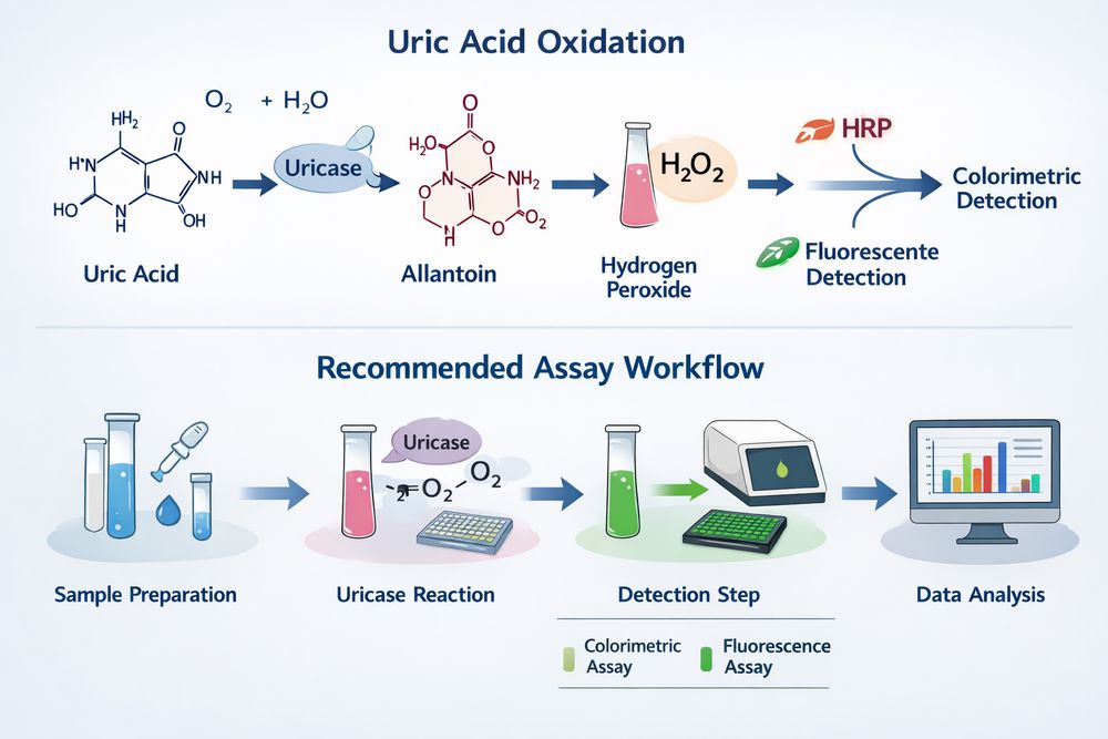
Fig 1. Reaction overview for uric acid oxidation and recommended assay workflow
1. Principle of the Uricase Activity Assay
The most commonly used assay format relies on the characteristic ultraviolet absorbance of uric acid at 293 nm. As uricase converts uric acid into downstream products, absorbance at 293 nm decreases in proportion to enzyme activity, allowing continuous kinetic monitoring.
Alternative analytical strategies include coupled assays that quantify hydrogen peroxide generation, chromatographic methods that resolve substrate and products simultaneously, and fluorometric systems that extend sensitivity. Method selection should be based on experimental goals, instrument availability, expected matrix interference, and required sensitivity.
1.1 Method Selection Guidance
- UV spectrophotometry: Best for routine QC, rapid kinetics, and straightforward purified-enzyme measurements.
- HPLC analysis: Best for mechanistic studies, product profiling, and complex biological samples.
- Coupled enzymatic assay: Best when higher sensitivity is required or when the direct UV method is limited by background interference.
For most research-grade pegloticase samples, begin with the 293 nm spectrophotometric assay, then confirm questionable or matrix-heavy samples by HPLC.
2. Required Reagents and Equipment
Accurate activity measurement depends on freshly prepared reagents, validated pH control, and stable instrumentation. The following list covers the standard requirements for both UV-based and HPLC-based uricase assays.
| Reagent | Specification | Purpose | Storage |
|---|---|---|---|
| Uric acid | ≥99% purity, crystalline | Primary substrate | -20°C, desiccated; prepare fresh solutions |
| Sodium phosphate buffer | 0.1 M, pH 7.4-8.5 | Reaction buffer | 4°C; verify pH before use |
| Potassium oxonate | ≥98% purity | Negative control inhibitor | -20°C |
| 4-Aminoantipyrine | Analytical grade | Chromogen for coupled assay | 4°C, protected from light |
| Phenol | Analytical grade | Chromogen component | 4°C |
| Peroxidase | Type II, ≥150 U/mg | Coupling enzyme | -20°C |
| Allantoin standard | ≥98% purity | HPLC product calibration | Room temperature, dry |
| Acetonitrile | HPLC grade | Mobile phase component | Room temperature |
| Formic acid | LC-MS grade | Mobile phase modifier | Room temperature |
2.1 Equipment Checklist
- UV-Vis spectrophotometer with kinetic mode and temperature control
- HPLC system with UV detector at 280-293 nm or MS detection
- 96-well microplate reader for high-throughput assays
- Water bath or equivalent temperature-equilibration device
- Analytical balance with 0.1 mg readability
- Calibrated pH meter, vortex mixer, microcentrifuge, and calibrated pipettes
3. Sample Preparation
Reliable activity data depend on careful sample handling, especially for PEGylated uricase preparations. Pegloticase has a high apparent molecular weight and should be handled gently to avoid foaming, adsorption loss, or inaccurate dispensing.
3.1 Enzyme Stock Preparation
3.2 Substrate Preparation
- Dissolve uric acid in a minimal volume of 0.1 M NaOH.
- Adjust the solution to pH 7.4-8.5 with phosphate buffer.
- Confirm full dissolution visually; turbidity suggests supersaturation or incomplete solubilization.
- Prepare uric acid fresh each day and avoid storage beyond 24 hours.
3.3 Biological Sample Processing
- Cell culture supernatant: Centrifuge at 10,000 × g for 10 minutes and assay immediately or store at -80°C.
- Tissue homogenate: Homogenize in 3-5 volumes of cold assay buffer, centrifuge at 12,000 × g for 15 minutes at 4°C, and collect the supernatant.
- Plasma or serum: Collect in heparin or EDTA tubes, centrifuge promptly, and process without delay. Freeze for extended storage.
3.4 Protein Normalization
- BCA assay: Generally compatible with PEGylated proteins.
- Bradford assay: Validate first because PEG can interfere with signal response.
- UV absorbance: Use A280 with an appropriate extinction coefficient when available.
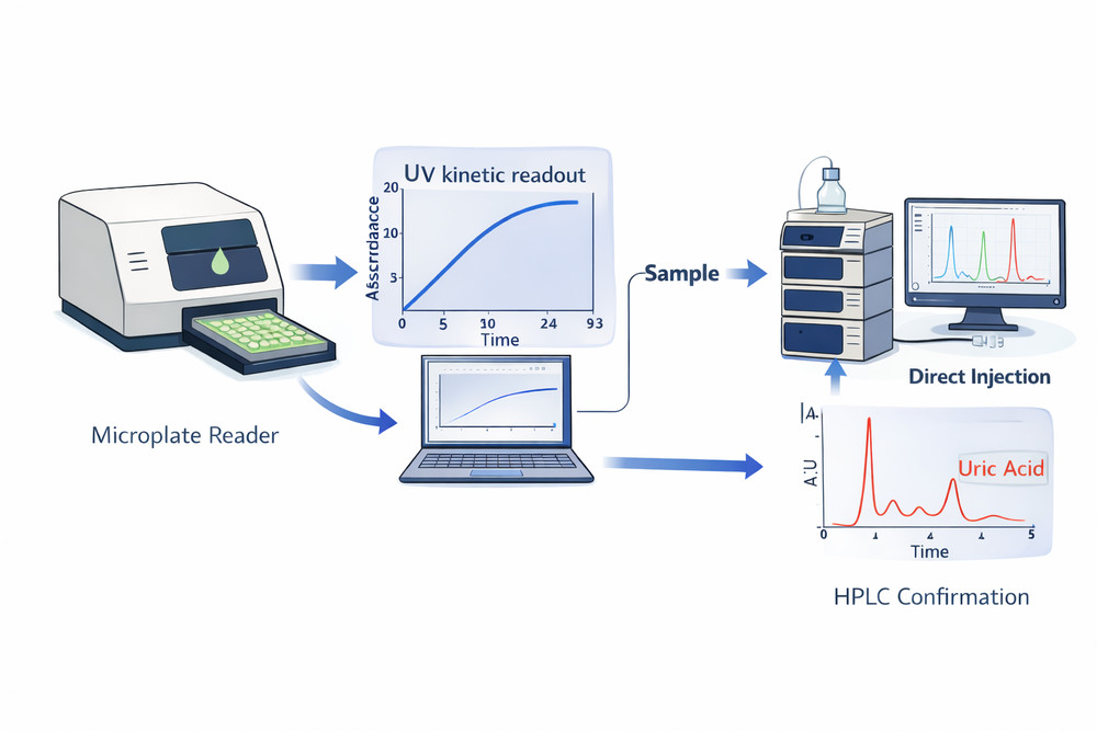
Fig 2. Suggested visual for assay setup, including UV kinetic readout and HPLC confirmation workflow
4. Reaction Setup and Standard Conditions
Standardized reaction conditions are essential for reproducibility across analysts, instruments, and laboratories. The table below summarizes the preferred operating range for routine uricase testing.
| Parameter | Standard Condition | Rationale | Acceptable Range |
|---|---|---|---|
| Temperature | 37°C | Physiological relevance | 25-40°C; maintain within ±0.5°C |
| pH | 7.4-8.5 | Optimal uricase activity window | 7.0-9.0 |
| Uric acid concentration | 100-600 μM | Linear range and saturation coverage | 50-1000 μM |
| Enzyme concentration | 0.1-10 μg/mL | Linear response range | Adjust to sample activity |
| Reaction volume | 1 mL cuvette or 200 μL microplate | Standard analytical format | Scale proportionally |
| Path length | 1 cm | Standard optical assumption | Correct for microplate geometry |
4.1 Spectrophotometric Assay Procedure
4.2 Coupled Colorimetric Assay
For greater sensitivity or for samples with UV interference, use a peroxidase-coupled system containing borate buffer, uric acid, 4-aminoantipyrine, phenol, and excess peroxidase. After incubation at 37°C, terminate the reaction and read absorbance at 540 nm against appropriate blanks.
4.3 Microplate Format
For high-throughput studies, dispense 180 μL of substrate solution into each well, pre-equilibrate the plate, add 20 μL of enzyme sample, mix for 10 seconds, and collect 293 nm data at 30-60 second intervals for 10 minutes. Apply path-length correction as required by the instrument and plate geometry.
5. Detection Methods
The analytical readout selected for uricase activity measurement will directly affect sensitivity, specificity, and throughput. Three common strategies are outlined below.
5.1 UV Spectrophotometry
- Principle: Uric acid absorbs strongly at 293 nm, with a molar extinction coefficient of 12,200 M-1cm-1.
- Advantages: Simple setup, real-time kinetic monitoring, and no coupling reagents required.
- Limitations: Moderate sensitivity and possible interference from UV-absorbing sample components.
- Typical sensitivity: Approximately 5-10 μM.
5.2 HPLC Analysis
HPLC provides improved specificity and can simultaneously quantify uric acid, 5-hydroxyisourate, and allantoin. A typical setup uses a C18 reverse-phase column, 5-10% acetonitrile in 0.1% formic acid, a 0.5-1.0 mL/min flow rate, and UV detection at 280-293 nm for uric acid or 220 nm for allantoin.
- Recommended injection volume: 10-20 μL
- Typical runtime: 10-15 minutes
- Best use case: mechanistic studies and complex sample matrices
5.3 Coupled Enzymatic Readout
This indirect approach measures hydrogen peroxide generated during uric acid oxidation. The peroxide signal is converted into a colored or fluorescent reporter using peroxidase, 4-aminoantipyrine, and phenol.
H2O2 + 4-aminoantipyrine + phenol → Quinoneimine dye + H2O
This format is especially helpful when direct UV detection is limited by sample background, but it requires excess peroxidase and careful control of antioxidant interference.
6. Data Analysis and Normalization
Convert raw absorbance or chromatographic values into meaningful activity units using a consistent and validated calculation workflow.
6.1 Activity Calculation
Specific Activity (U/mg) = [Rate × Reaction Volume (L)] / Enzyme Mass (mg)
One unit of uricase activity is defined as the amount of enzyme that oxidizes 1 μmol of uric acid per minute under standard assay conditions.
6.2 Normalization Strategies
| Normalization Basis | Application | Calculation Format | Considerations |
|---|---|---|---|
| Total protein | Purified enzyme preparations | Activity/mg protein | Use an accurate protein assay and account for PEG effects |
| Cell number | Cell culture studies | Activity/106 cells | Validate cell counting and lysis completeness |
| Tissue weight | Tissue homogenates | Activity/g wet tissue | Standardize homogenization and note blood contamination |
| Volume | Biological fluids | Activity/mL | Report dilution factors and matrix effects |
6.3 Kinetic Characterization
For mechanistic studies, measure initial rates across a substrate range of 10-500 μM uric acid, fit the data to the Michaelis-Menten model, and report Km, Vmax, and catalytic efficiency. Typical pegloticase values fall around Km ≈ 10-50 μM and kcat ≈ 10-100 s-1, depending on PEGylation extent and assay conditions.
7. Troubleshooting Tips
Common assay failures usually stem from enzyme instability, incomplete uric acid dissolution, temperature drift, or optical interference. Use the table below to diagnose routine issues quickly.
| Problem | Possible Cause | Recommended Solution |
|---|---|---|
| No activity detected | Enzyme denaturation, substrate precipitation, or incorrect wavelength | Prepare fresh enzyme, verify uric acid dissolution, and confirm the instrument is set to 293 nm |
| High background absorbance | Contaminated buffer, impure substrate, or sample interference | Use fresh reagents, improve substrate purity, or switch to HPLC for complex matrices |
| Nonlinear kinetics | Substrate depletion, product inhibition, or enzyme instability | Measure only the initial rate, reduce enzyme load, or increase substrate concentration |
| High variability | Pipetting error, temperature fluctuation, or incomplete mixing | Calibrate pipettes, confirm thermal stability, and improve mixing consistency |
| Low sensitivity | Instrument limitation or suboptimal pH | Inspect optics, verify lamp performance, and optimize the buffer within the pH 7.4-8.5 range |
Include a positive control in every run, define inter-assay acceptance criteria such as CV <15%, confirm dilution linearity, and monitor standard curves or system suitability over time.
For applications requiring a stable and well-characterized enzyme source, Pegloticase, Recombinant Uricase can support reproducible assay performance across extended experimental workflows.