Using Human Hyaluronidase in Extracellular Matrix and Tissue Permeability Research
A Practical Framework for ECM Remodeling, Drug Penetration Models, and Quantitative Permeability Assays
Abstract
The extracellular matrix (ECM) presents a significant physical barrier to drug delivery, cell migration, and molecular diffusion in tissue models. Human hyaluronidase specifically degrades hyaluronan (HA), the primary glycosaminoglycan responsible for ECM viscosity and pore size regulation. This application note provides a practical framework for using recombinant human hyaluronidase (PH20) in three experimental contexts: gel-based matrix models, 3D cell culture systems, and ex vivo tissue permeability assays. We outline critical experimental variables, quantitative readouts, and interpretation guidelines to help researchers generate reproducible, biologically meaningful data while acknowledging the inherent limitations of in vitro ECM surrogates.
human hyaluronidase extracellular matrix, tissue permeability, ECM remodeling, hyaluronan, drug penetration models, 3D cell culture
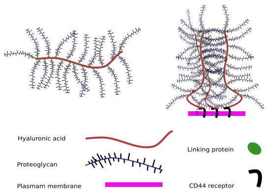
Fig 1. Hyaluronan forms a hydrated meshwork in the ECM by interacting with proteoglycans and link proteins, creating a viscoelastic barrier that regulates molecular diffusion and cell migration.
1. Why Hyaluronan Matters in ECM Structure
Hyaluronan (also called hyaluronic acid or hyaluronate) is a non-sulfated, linear glycosaminoglycan composed of repeating disaccharide units of N-acetylglucosamine and glucuronic acid. Unlike other ECM components, HA is not covalently bound to a protein core; instead, it exists as a free polysaccharide that interacts with proteoglycans, link proteins, and cell surface receptors such as CD44 and RHAMM. These interactions create a hydrated, viscoelastic meshwork that occupies the interstitial spaces of connective tissues.
The biological significance of HA in ECM architecture stems from three unique physicochemical properties:
- Extraordinary hydration capacity: HA can bind up to 1,000 times its weight in water, generating osmotic pressure that maintains tissue turgor and creates a hydrated microenvironment for cell signaling.
- Length-dependent pore size regulation: High-molecular-weight HA (HMW-HA, >1,000 kDa) forms entangled networks with small mesh sizes (tens of nanometers), physically excluding large molecules and creating a diffusion barrier.
- Dynamic turnover: HA is continuously synthesized by hyaluronan synthases (HAS1–3) and degraded by hyaluronidases (HYAL1–4) and reactive oxygen species, allowing rapid ECM remodeling in response to injury, inflammation, or disease.
In pathological states such as solid tumors, fibrosis, and chronic inflammation, HA deposition is often markedly elevated. Tumor-associated stroma, for example, can contain 5- to 20-fold higher HA concentrations than normal tissue, contributing to elevated interstitial fluid pressure (IFP) and impaired drug penetration. This makes HA a rational target for enzymatic modulation in permeability research.
2. Human Hyaluronidase as an ECM-Modulating Enzyme
Recombinant human hyaluronidase (rHuPH20) is a soluble, neutral-active enzyme expressed in CHO cells that catalyzes the hydrolysis of the β-1,4-glycosidic bond between glucuronic acid and N-acetylglucosamine residues in HA. The enzyme exhibits optimal activity at pH 5.7–7.0 and 37°C, making it compatible with standard cell culture conditions and physiological tissue environments.
2.1 Mechanism of Action
rHuPH20 operates through an endo-β-N-acetylhexosaminidase mechanism, randomly cleaving internal glycosidic bonds within the HA polymer chain. This process rapidly reduces the molecular weight and chain length of HA, which in turn:
- Decreases solution viscosity (η) by disrupting polymer entanglement
- Increases the average mesh size (ξ) of the ECM network
- Reduces osmotic pressure by releasing bound water molecules
- Generates HA oligosaccharides that may act as signaling molecules
Enzymatic digestion of HA by rHuPH20 does not simply "dissolve" the matrix; it remodels the pore architecture in a concentration- and time-dependent manner, enabling tunable permeability control in experimental models.
2.2 Product Specifications for Research Use
| Attribute | Specification | Relevance to ECM Research |
|---|---|---|
| Source | CHO cell expression system | Mammalian glycosylation ensures proper folding and activity |
| Molecular weight | ~61 kDa | Small enough for uniform distribution in gel matrices |
| Specific activity | 6.5–400 KU/mL | Wide dosing range for matrix density optimization |
| Endotoxin level | <0.1 EU/μg protein | Minimizes inflammatory confounders in cell-based assays |
| Optimal pH | 5.7–7.0 | Compatible with standard culture media (pH 7.2–7.4) |
| Purity | >95% (SDS-PAGE) | Reduces off-target proteolytic activity |
One unit of rHuPH20 activity is defined as the amount of enzyme that causes a change in absorbance at 600 nm of 0.330 per minute at pH 5.7 and 37°C using a turbidimetric assay with HA substrate. Researchers should verify unit consistency when comparing data across suppliers, as assay conditions may vary.
3. Tissue Permeability Models
Three principal model systems are used to study hyaluronidase-mediated ECM remodeling and tissue permeability. Each offers distinct advantages and limitations, and the choice depends on the research question, throughput requirements, and desired biological complexity.
3.1 Gel Matrix Models
Gel-based matrices—most commonly hyaluronan-based hydrogels, Matrigel, or collagen I gels—provide the simplest and most controllable environment for studying enzyme-mediated permeability changes.
Typical workflow:
- Prepare HA hydrogels at defined concentrations (e.g., 1–5 mg/mL) and crosslinking densities.
- Load a fluorescent tracer (e.g., FITC-dextran of known molecular weight, 4–500 kDa) or a model drug into a donor chamber.
- Apply rHuPH20 at varying concentrations (10–1,000 U/mL) to the gel surface or mix into the gel precursor.
- Incubate at 37°C for defined time intervals (15 min to 24 h).
- Quantify tracer diffusion into a receiver compartment or penetration depth via confocal microscopy.
Advantages: High reproducibility, precise control over matrix composition, amenable to high-throughput screening, and minimal ethical constraints.
Limitations: Lack of cellular components, absence of active ECM remodeling, and simplified mechanical properties compared to native tissue.
3.2 3D Cell Culture Models
Three-dimensional cell cultures—such as tumor spheroids, organoids, or fibroblast-populated collagen lattices—introduce living cells that actively synthesize and remodel ECM, providing a more physiologically relevant context.
In these models, rHuPH20 is typically applied to established 3D structures after a maturation period (3–14 days) to allow endogenous ECM deposition. The enzyme can be delivered as a bolus, in sustained-release formulations, or conjugated to nanoparticles for localized matrix remodeling.
When using rHuPH20 in 3D cultures, enzyme exposure time must be balanced against cell viability. Prolonged incubation (>24 h) at high concentrations (>500 U/mL) may reduce cell viability in some spheroid models, likely due to HA fragment-mediated signaling rather than direct cytotoxicity. Include appropriate vehicle controls and viability assays (e.g., CellTiter-Glo, LIVE/DEAD staining).
3.3 Ex Vivo Tissue Models
Ex vivo tissue slices—such as human skin explants, tumor xenograft sections, or resected organ tissue—retain native ECM architecture, cellular heterogeneity, and vascular structures, offering the highest biological fidelity among in vitro approaches.
| Model Type | Biological Complexity | Throughput | Reproducibility | Best Application |
|---|---|---|---|---|
| Gel matrix | Low | High (96/384-well) | Excellent (CV <10%) | Mechanism screening, dose optimization |
| 3D cell culture | Medium | Medium (24/96-well) | Good (CV 10–20%) | Cell-ECM interaction studies |
| Ex vivo tissue | High | Low (6/12-well) | Moderate (CV 15–30%) | Translational validation, clinical relevance |
Fig 2. Schematic representation of hyaluronidase-mediated ECM barrier reduction. Enzymatic digestion of HA decreases matrix density, enlarging pore size and facilitating molecular penetration through the interstitial space.
4. Experimental Variables
Reproducible permeability data requires rigorous control of three interdependent variables: enzyme concentration, exposure time, and matrix density. Each parameter nonlinearly influences the others, and their optimization is assay-specific.
4.1 Enzyme Concentration
The relationship between rHuPH20 concentration and HA degradation follows Michaelis-Menten kinetics at subsaturating substrate concentrations, but deviates at high enzyme doses due to substrate depletion and product inhibition. Typical working concentrations range from 10 U/mL (subtle matrix softening) to 1,000 U/mL (near-complete HA digestion).
Recommended dose-escalation strategy:
- Pilot study: 10, 50, 100, 500, 1,000 U/mL
- Confirmatory study: Narrow around the EC₅₀ for your specific matrix
- Always include a heat-inactivated enzyme control to rule out non-enzymatic effects
4.2 Exposure Time
HA degradation kinetics are biphasic: an initial rapid phase (0–2 h) during which HMW-HA is cleaved into intermediate fragments, followed by a slower phase (2–24 h) of further oligosaccharide generation. For most permeability assays, a 2- to 4-hour incubation captures the maximal change in diffusion properties without excessive HA depletion.
4.3 Matrix Density
Matrix density—whether defined by HA concentration, collagen concentration, or total protein content—determines the baseline pore size and thus the magnitude of hyaluronidase effect. Dense matrices (e.g., 5 mg/mL HA or 4 mg/mL collagen) show larger relative permeability increases upon HA digestion compared to sparse matrices, but also require higher enzyme doses to achieve comparable fractional degradation.
| Variable | Typical Range | Impact on Readout | Optimization Tip |
|---|---|---|---|
| Enzyme concentration | 10–1,000 U/mL | Dose-dependent viscosity reduction; plateau at saturation | Use log-scale dilutions; verify activity with standard curve |
| Exposure time | 15 min – 24 h | Biphasic kinetics; rapid initial phase, slower secondary phase | Time-course pilot (0.5, 1, 2, 4, 8, 24 h) |
| Matrix density | 1–10 mg/mL (HA) 1–6 mg/mL (collagen) |
Higher density = larger relative effect but requires more enzyme | Match density to target tissue (tumor stroma ~3–5 mg/mL HA) |
| Temperature | 25–37°C | Activity increases ~2-fold per 10°C (Q₁₀ ≈ 2) | Maintain 37°C ± 0.5°C for physiological relevance |
| pH | 6.0–7.5 | Optimal at pH 5.7–7.0; activity drops >50% at pH >7.5 | Use buffered media (PBS or DMEM); monitor CO₂ in incubators |
5. Readouts
Quantitative readouts for hyaluronidase-mediated permeability fall into four categories: bulk diffusion metrics, spatial penetration profiles, rheological properties, and imaging-based structural analysis.
5.1 Diffusion Rate
Diffusion coefficients (D) can be measured using:
- Franz diffusion cells: Measure cumulative permeation of a tracer across a tissue or gel membrane over time. Calculate flux (J) and permeability coefficient (Kp).
- Fluorescence recovery after photobleaching (FRAP): Determine local diffusion coefficients within the matrix by photobleaching a small region and measuring fluorescence recovery kinetics.
- Dynamic light scattering (DLS): Track changes in tracer hydrodynamic radius or matrix mesh size indirectly.
5.2 Penetration Depth
Confocal or multiphoton microscopy enables direct visualization of fluorescent tracer penetration from the matrix surface. Penetration depth (δ) is typically defined as the distance at which fluorescence intensity drops to 1/e (37%) of the surface value. rHuPH20 treatment can increase δ by 2- to 5-fold in dense HA gels, depending on enzyme dose and tracer size.
5.3 Viscosity Change
Rheological measurements provide bulk mechanical readouts of matrix remodeling:
| Rheological Parameter | Measurement | Expected Change with rHuPH20 | Interpretation |
|---|---|---|---|
| Storage modulus (G′) | Oscillatory shear rheometry | Decrease 30–80% | Reduced elastic network integrity |
| Loss modulus (G″) | Oscillatory shear rheometry | Decrease 20–60% | Reduced viscous dissipation |
| Complex viscosity (η*) | Steady shear or oscillation | Decrease 40–90% | Overall matrix fluidization |
| Tan δ (G″/G′) | Frequency sweep | Increase (approach 1.0) | Shift toward liquid-like behavior |
5.4 Imaging
Advanced imaging techniques provide structural context for permeability changes:
- Second harmonic generation (SHG) microscopy: Visualizes collagen fiber architecture and alignment changes following HA removal.
- Transmission electron microscopy (TEM): Resolves nanoscale pore structure and HA filament density at high resolution.
- Magnetic resonance imaging (MRI): Maps T2 relaxation time changes associated with water content redistribution in larger tissue samples.
6. Interpreting Results Without Overstating Biological Relevance
A common pitfall in ECM permeability research is extrapolating in vitro findings directly to in vivo drug delivery outcomes. While rHuPH20 clearly increases diffusion in gel and tissue models, several factors moderate translational relevance:
- Enzyme distribution in vivo: In a living tissue, rHuPH20 is subject to convective clearance, proteolytic degradation, and binding to endogenous inhibitors—factors absent in static gel models.
- Compensatory ECM synthesis: Cells in vivo upregulate HAS expression in response to HA depletion, partially restoring matrix density within hours to days.
- Heterogeneous HA distribution: Native tissues exhibit spatially variable HA density (e.g., pericellular vs. interstitial), whereas model matrices are typically homogeneous.
- Immunogenicity concerns: Repeated administration of non-human or even human recombinant enzymes can elicit antibody responses in vivo, limiting long-term efficacy.
Frame in vitro permeability data as "proof of mechanism" or "barrier reduction potential" rather than "predicted clinical efficacy." Use phrases such as "rHuPH20 increased diffusion coefficient by 3.2-fold in this model" rather than "rHuPH20 improves drug delivery."
When reporting results, clearly distinguish between:
- Matrix-level effects: Changes in pore size, viscosity, or diffusion coefficient directly attributable to HA degradation.
- Cell-level effects: Changes in cell behavior (migration, proliferation, signaling) that may result from HA fragment generation or mechanical cues.
- Tissue-level effects: Integrated responses in complex models (e.g., spheroid drug penetration) that reflect both matrix and cellular contributions.
7. Limitations of In Vitro ECM Models
All model systems simplify the native ECM. Understanding these limitations is essential for appropriate experimental design and data interpretation.
7.1 Composition Simplification
Native ECM contains hundreds of distinct molecules—collagens, elastin, fibronectin, laminin, proteoglycans, and glycoproteins—arranged in tissue-specific architectures. Most in vitro models use one or two purified components (e.g., HA + collagen I), missing critical interactions that regulate permeability in vivo.
7.2 Absence of Cellular Turnover
Static gel models lack living cells that continuously synthesize, degrade, and reorganize ECM. Even 3D cultures often fail to replicate the dynamic balance between synthesis and degradation observed in vivo, particularly over extended culture periods (>2 weeks).
7.3 Mechanical Mismatch
The stiffness of common gel matrices (0.1–10 kPa) overlaps with soft tissues but may not match the mechanical properties of target tissues such as desmoplastic tumors (>20 kPa) or cartilage (>100 kPa). Mechanical mismatch can alter cell behavior and ECM deposition patterns.
7.4 Scale and Geometry
Most in vitro models use planar geometries (Transwell inserts, gel slabs) with thicknesses of 100–500 μm. Native tissue barriers—such as tumor stroma or dermis—span millimeters to centimeters, introducing transport phenomena (convection, pressure gradients) not captured in thin models.
To improve translational relevance: (1) use patient-derived xenograft (PDX) tissue when possible; (2) incorporate physiological flow (microfluidic devices) to simulate interstitial fluid pressure; (3) match matrix stiffness to target tissue using tunable crosslinkers; and (4) validate key findings in at least two model systems of differing complexity.
8. Study Design Checklist
Use this checklist to ensure rigorous experimental design and reporting for hyaluronidase-mediated permeability studies.
Pre-Experimental Planning
- Define primary research question: mechanism, optimization, or translational validation?
- Select model system appropriate to biological complexity required (gel → 3D culture → ex vivo).
- Match matrix composition and density to target tissue literature values.
- Calculate required sample size based on expected effect size and pilot data variability.
Experimental Execution
- Include vehicle control (buffer only), heat-inactivated enzyme control, and positive control (if available).
- Randomize sample placement in multi-well plates to avoid position effects.
- Run experiments in at least biological triplicate (independent gel/culture preparations).
- Document enzyme batch number, specific activity, and storage conditions.
- Maintain constant temperature (37°C) and pH (7.2–7.4) throughout incubation.
Data Analysis and Reporting
- Report raw data (not just fold-change) with appropriate units (cm²/s for D, μm for δ, Pa·s for η).
- Use appropriate statistical tests; account for repeated measures if sampling the same gel over time.
- Report limitations explicitly, including model simplifications and potential confounders.
- Avoid causal language for correlational data; use "associated with" rather than "causes."
- Deposit raw data and analysis code in public repositories when possible.
References
1. Toole, B. P. (2004). Hyaluronan: from extracellular glue to pericellular cue. Nature Reviews Cancer, 4(7), 528-539.
2. Stern, R., Asari, A. A., & Sugahara, K. N. (2006). Hyaluronan fragments: an information-rich system. European Journal of Cell Biology, 85(8), 699-715.
3. Bookbinder, L. H., et al. (2006). A recombinant human enzyme for enhanced interstitial transport of therapeutics. Journal of Controlled Release, 114(2), 230-241.
4. Thorne, R. G., & Nicholson, C. (2006). Role of tortuosity in anomalous diffusion in the brain. Biophysical Journal, 91(8), 2965-2970.
5. Netti, P. A., et al. (2000). Role of extracellular matrix assembly in interstitial diffusion in normal and tumor tissue. Cancer Research, 60(9), 2497-2503.
6. Kultti, A., et al. (2012). Therapeutic targeting of hyaluronan in the tumor stroma. Cancers, 4(3), 873-903.
7. Provenzano, P. P., et al. (2012). Enzymatic targeting of the stroma ablates physical barriers to treatment of pancreatic ductal adenocarcinoma. Cancer Cell, 21(3), 418-429.
8. Chauhan, V. P., et al. (2013). Angiotensin inhibition enhances drug delivery and potentiates chemotherapy by decompressing tumour blood vessels. Nature Communications, 4, 2516.
9. Jain, R. K., & Stylianopoulos, T. (2010). Delivering nanomedicine to solid tumors. Nature Reviews Clinical Oncology, 7(11), 653-664.
10. Frampton, J. E. (2010). Hyaluronidase (rHuPH20): an enabling technology for subcutaneous drug delivery. BioDrugs, 24(5), 295-299.