Human Hyaluronidase FAQ
Mechanism, Activity Units, Storage, and Research Use
Basic Concepts
Human hyaluronidase is a family of enzymes that catalyze the hydrolysis of hyaluronan (also known as hyaluronic acid or HA), a high-molecular-weight glycosaminoglycan abundant in the extracellular matrix. By cleaving the β-1,4-glycosidic bonds between N-acetylglucosamine and D-glucuronic acid residues, human hyaluronidase reduces the viscosity of connective tissue and increases tissue permeability. In clinical and research settings, this enzyme serves as a spreading factor that facilitates the dispersion and absorption of co-injected substances.
Native human hyaluronidases are classified into two main groups: HYAL1 (lysosomal, active at acidic pH) and PH20 (sperm-associated, active at neutral pH). The recombinant form used in most research and therapeutic applications is derived from the PH20 isoform due to its optimal activity at physiological pH.
Recombinant human hyaluronidase PH20 is a soluble fragment of the human sperm surface protein PH20, expressed in Chinese Hamster Ovary (CHO) cells. The purified glycoprotein contains 447 amino acids with an approximate molecular weight of 61,000 Daltons. Unlike native PH20, which is a glycosylphosphatidylinositol (GPI)-anchored membrane protein, the recombinant variant lacks the transmembrane domain and is secreted as a soluble enzyme.
Expression in CHO cells ensures proper mammalian glycosylation patterns, which are essential for enzymatic activity and stability. The recombinant production system eliminates risks associated with animal-derived materials and provides batch-to-batch consistency critical for research reproducibility.
Mechanism and Activity
The catalytic mechanism of PH20 involves hydrolytic cleavage of the β-1,4-glycosidic linkages within the hyaluronan polymer chain. The enzyme operates through an acid-base catalysis mechanism involving two conserved glutamate residues in the active site:
- Substrate binding: The enzyme recognizes the repeating disaccharide units of hyaluronan (GlcNAc-β-1,4-GlcA) through a deep catalytic cleft.
- Hydrolysis: A general acid (Glu residue) protonates the glycosidic oxygen, while a general base activates a water molecule for nucleophilic attack at the anomeric carbon.
- Product release: Cleavage generates smaller oligosaccharide fragments (tetrasaccharides and hexasaccharides) that diffuse away from the active site.
At physiological pH (7.4), PH20 exhibits optimal activity, whereas HYAL1 requires acidic conditions (pH 3.5-4.5) found in lysosomes. This pH profile makes PH20 the preferred enzyme for in vivo and cell-based applications.
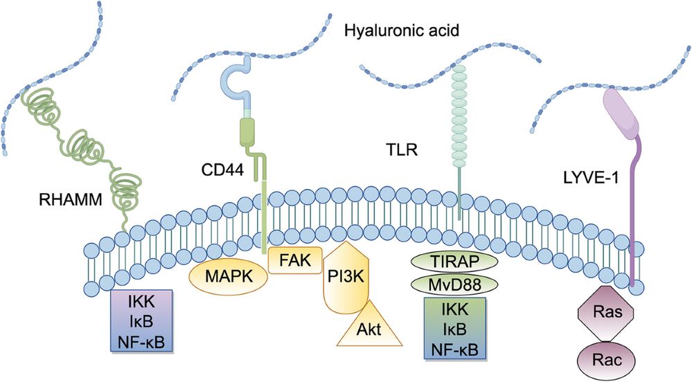
Fig 1. Hyaluronidase structure, mechanism of action, and cellular signaling pathways involving hyaluronan degradation
Hyaluronidase activity is quantified using a turbidimetric assay based on the method originally described by Dorfman and Ott (1948). One unit (U) of recombinant human hyaluronidase activity is defined as the amount of enzyme that causes a change in absorbance at 600 nm (A600nm) of 0.330 per minute under standard assay conditions: pH 5.7, 37°C, using hyaluronan as substrate.
The assay principle relies on the precipitation of undigested hyaluronan with acidified serum albumin. As the enzyme degrades hyaluronan into smaller fragments, fewer large polymers remain available for precipitation, resulting in decreased turbidity. The rate of turbidity reduction is directly proportional to enzyme activity.
| Parameter | Standard Condition | Optimal Range |
|---|---|---|
| pH | 5.7 | 5.5 - 6.0 |
| Temperature | 37°C | 35 - 40°C |
| Substrate | Hyaluronan (0.5 mg/ml) | 0.3 - 1.0 mg/ml |
| Ionic Strength | 0.1 M NaCl | 0.05 - 0.2 M |
| Detection Wavelength | 600 nm | 580 - 640 nm |
| Unit Definition | ΔA600nm = 0.330/min | |
Activity measurements are highly sensitive to assay conditions. Variations in pH, ionic strength, and substrate concentration can significantly affect reported unit values. Always verify the assay protocol used by the supplier when comparing products from different sources.
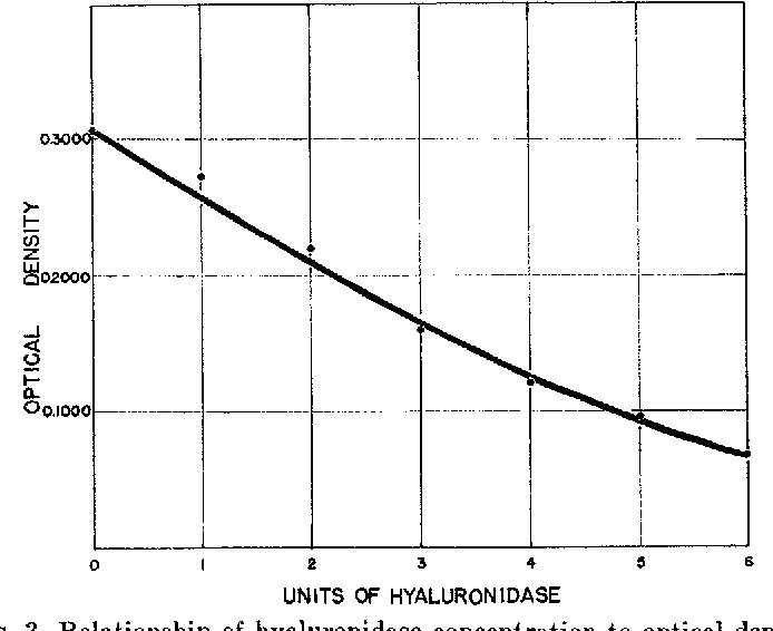
Fig 2. Standard curve showing the relationship between hyaluronidase concentration and optical density in the turbidimetric assay
Expression and Quality
The expression system significantly impacts the functional properties of recombinant human hyaluronidase due to differences in post-translational modifications, particularly glycosylation:
| Feature | CHO Cells (Mammalian) | Yeast (Pichia) | E. coli (Bacterial) |
|---|---|---|---|
| Glycosylation | Complex, human-like | High-mannose | None |
| Enzymatic Activity | High (native conformation) | Moderate | Low/Variable |
| Endotoxin Risk | Very Low | Low | High |
| Batch Consistency | Excellent | Good | Moderate |
| Production Cost | Higher | Moderate | Lower |
| Research Suitability | Optimal | Acceptable | Limited |
CHO cell expression is the gold standard for human hyaluronidase production because:
- Proper N-linked glycosylation at six consensus sites is required for enzymatic activity and thermal stability
- Absence of immunogenic non-human glycan epitopes (e.g., α-Gal, Neu5Gc)
- Ability to produce the correct disulfide bond pattern (8 cysteine residues)
- Regulatory acceptance for therapeutic-grade materials
Storage and Handling
Proper PH20 storage is critical for maintaining enzymatic activity and preventing aggregation:
| Storage Condition | Temperature | Expected Stability | Notes |
|---|---|---|---|
| Long-term | -80°C | ≥ 24 months | Preferred for stock solutions |
| Short-term | -20°C | 6 - 12 months | Avoid repeated freeze-thaw |
| Working aliquots | 4°C | 1 - 2 weeks | Keep sterile, avoid light |
| Avoid | Room temp | < 24 hours | Rapid activity loss |
Best Practices for Handling:
- Aliquot into single-use volumes immediately upon receipt to minimize freeze-thaw cycles
- Use low-protein-binding tubes (polypropylene) for storage
- Maintain pH between 6.0 - 7.5 in buffer systems (PBS, HEPES, or Tris-based)
- Include stabilizers such as 0.1% BSA or 10% glycerol for dilute solutions (< 0.1 mg/ml)
- Avoid vortexing; gentle mixing by inversion prevents mechanical denaturation
- Protect from light if the formulation contains photosensitive excipients
Recombinant human hyaluronidase is sensitive to freeze-thaw stress. Each freeze-thaw cycle can reduce activity by 5-15%. Always prepare working aliquots and thaw rapidly in a 37°C water bath with gentle agitation, then immediately place on ice for use.
Experimental Applications
Yes, recombinant human hyaluronidase is widely used in cell-based assays, but several considerations ensure valid results:
Common Cell-Based Applications:
- 3D spheroid/tumoroid dissociation: Enzymatic digestion of hyaluronan-rich matrices to release single cells for flow cytometry or subculture
- Cell migration studies: Modulation of extracellular matrix barrier properties to assess invasive potential
- Drug penetration assays: Co-treatment to enhance therapeutic antibody or nanoparticle penetration in tumor models
- Immunohistochemistry: Tissue permeabilization to improve antibody penetration in thick sections
Cell Compatibility Considerations:
- Endotoxin levels must be < 0.1 EU/μg to avoid TLR4-mediated inflammatory responses in immune cells
- Concentrations typically range from 10 - 100 U/ml depending on the matrix density
- Incubation time should be optimized (usually 15 - 60 minutes at 37°C)
- Some cell types (e.g., chondrocytes, mesenchymal stem cells) are sensitive to hyaluronan depletion; include recovery periods post-treatment
Rigorous experimental controls are essential for interpreting hyaluronidase results accurately:
| Control Type | Purpose | Implementation |
|---|---|---|
| Vehicle control | Account for buffer effects | Same buffer without enzyme |
| Heat-inactivated enzyme | Confirm specificity | 65°C for 30 min or boiling |
| Hyaluronan rescue | Verify substrate dependence | Re-add exogenous HA (100 μg/ml) |
| Positive control | Validate assay sensitivity | Known active enzyme batch |
| Dose-response | Establish linear range | Serial dilution (0.1 - 100 U/ml) |
| Time course | Determine kinetics | Sampling at 0, 15, 30, 60, 120 min |
For mechanistic studies, consider using specific hyaluronan synthase inhibitors (e.g., 4-MU) or competing substrates (chondroitin sulfate) to distinguish hyaluronan-specific effects from general matrix disruption.
Troubleshooting and Reporting
Unexpected loss of recombinant human hyaluronidase activity can usually be traced to the following factors:
Storage-Related Issues:
- Repeated freeze-thaw cycles causing protein aggregation
- Extended storage at 4°C leading to microbial contamination or proteolysis
- Exposure to extreme pH (< 4.0 or > 9.0) during buffer preparation
Assay-Related Issues:
- Incorrect pH: PH20 requires neutral pH (6.5 - 7.5); acidic conditions inactivate the enzyme
- Substrate quality: Hyaluronan molecular weight and purity affect reaction kinetics
- Inhibitors: Heavy metals (Hg²⁺, Cu²⁺), sulfated glycosaminoglycans, and certain dyes can inhibit activity
- Temperature deviations: Activity decreases by approximately 50% at 25°C compared to 37°C
Formulation Issues:
- Protein concentration too low (< 0.01 mg/ml) leading to surface adsorption losses
- Absence of stabilizing proteins (BSA) or sugars (trehalose) in dilute preparations
- Aggregation due to improper thawing techniques
Standardized reporting of human hyaluronidase experiments ensures reproducibility and comparability across studies:
Required Reporting Elements:
- Enzyme specifications: Source (CHO-derived), catalog number, lot number, stated activity (U/ml or U/mg), and endotoxin level
- Assay conditions: pH, temperature, substrate type and concentration, buffer composition, and reaction volume
- Unit definition: Explicit statement of the unit definition used (e.g., "Units as defined by turbidimetric assay at pH 5.7, 37°C")
- Activity calculation: Raw data (absorbance vs. time), standard curve parameters, and final activity with standard deviation
- Controls: Description of all controls used and their outcomes
"Recombinant human hyaluronidase PH20 (Cat# THP-0123, Lot# H2026001, 6.5 KU/ml, endotoxin <0.1 EU/μg) was diluted in PBS (pH 7.4) containing 0.1% BSA. Activity was confirmed by turbidimetric assay (ΔA600nm/min at pH 5.7, 37°C) using hyaluronan substrate (0.5 mg/ml). One unit corresponds to a change in A600nm of 0.330 per minute under these conditions."
References
1. Cohn, E. J., et al. (1946). J Am Chem Soc, 68(3): 459-475.
2. Dorfman, A., & Ott, M. L. (1948). J Biol Chem, 172(1): 367-375.
3. Frost, G. I. (2007). Expert Opin Drug Deliv, 4(4): 427-440.
4. Bookbinder, L. H., et al. (2006). J Control Release, 114(2): 230-241.
5. Stern, R. (2003). Eur J Cell Biol, 82(7): 345-349.
6. Csoka, A. B., et al. (2001). Matrix Biol, 20(8): 499-508.
7. European Pharmacopoeia 10.0, Hyaluronidase monograph.
8. United States Pharmacopeia (USP), Hyaluronidase Assay <91>.