How to Select Recombinant Human Hyaluronidase for Research
Source, Activity, Purity, and Endotoxin Considerations
1. Why Selection Criteria Matter
Recombinant human hyaluronidase PH20 is widely employed in biomedical research to degrade hyaluronic acid (HA)-rich extracellular matrices, facilitate drug dispersion, and modulate tumor microenvironments. However, not all commercial preparations are equivalent. Variations in recombinant human hyaluronidase products can lead to inconsistent enzymatic activity, unexpected cytotoxicity, or failed cell-based assays. This guide provides a systematic framework for evaluating hyaluronidase candidates across seven critical dimensions, enabling researchers to make data-driven purchasing decisions aligned with their experimental goals.
Enzyme source, glycosylation pattern, and endotoxin load are the three most common sources of assay variability in cell-based hyaluronidase studies. Standardizing these parameters improves inter-laboratory reproducibility.
2. Expression Source: CHO, HEK, and Microbial Systems
The choice of expression host profoundly affects the folding, glycosylation, and immunogenicity of recombinant human hyaluronidase PH20. Each platform presents distinct trade-offs between cost, scalability, and post-translational modification fidelity.
| Expression System | Glycosylation Profile | Advantages | Limitations | Best For |
|---|---|---|---|---|
| CHO (Chinese Hamster Ovary) | Complex, human-like N-glycans; sialylation present | Regulatory acceptance; scalable; authentic folding | Higher cost; longer production timelines | Preclinical pharmacology; long-term cell culture |
| HEK293 (Human Embryonic Kidney) | Human-identical glycosylation; α2-6 sialylation | Closest to native human PH20; high specific activity | Lower yields; higher per-mg cost | Mechanistic studies; protein interaction assays |
| Pichia pastoris (Yeast) | Hyper-mannosylated N-glycans; no sialylation | High expression titer; cost-effective; rapid turnaround | Non-human glycosylation may alter half-life and immunogenicity | Enzymatic screening; biochemical assays |
| E. coli (Bacterial) | No glycosylation; inclusion body risk | Lowest cost; fastest expression | Refolding required; often inactive or aggregated | Antigen production; non-glycosylated domain studies |
Research Implication: Mammalian systems (CHO or HEK) are strongly recommended for cell-based and in vivo applications where glycosylation-dependent stability and activity are critical. Microbial systems may suffice for simple turbidimetric activity assays or structural studies where glycan heterogeneity is not a confounding factor.
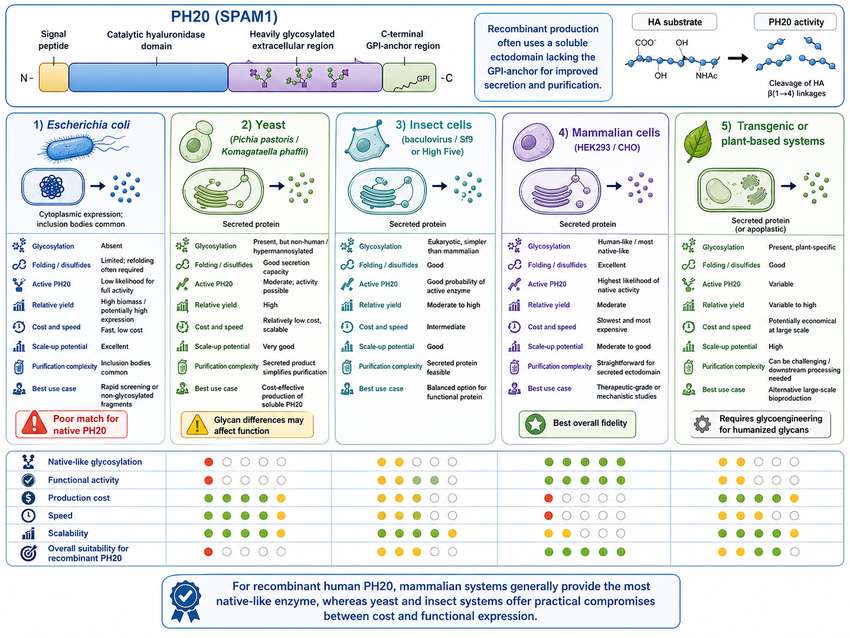
Fig 1. Comparative overview of expression system characteristics for recombinant human hyaluronidase PH20 production
3. Protein Identity: PH20 Sequence, Molecular Weight, and Glycosylation
3.1 Sequence Integrity
Native human PH20 is a glycosylphosphatidylinositol (GPI)-anchored protein with a molecular mass of approximately 64 kDa for the unglycosylated polypeptide. Research-grade hyaluronidase preparations should be verified by N-terminal sequencing or mass spectrometry to confirm:
- Full-length expression without unintended truncations
- Correct signal peptide cleavage
- Absence of frameshift mutations or amino acid substitutions
3.2 Molecular Weight and Glycosylation
SDS-PAGE under reducing conditions typically reveals a broad band between 60–75 kDa for mammalian-expressed PH20, reflecting heterogeneous N-glycosylation at multiple consensus sites (Asn-X-Ser/Thr). The extent of glycosylation directly impacts:
- Enzymatic activity: Properly glycosylated PH20 exhibits higher substrate turnover rates.
- Serum half-life: Sialylated glycans reduce clearance by hepatic asialoglycoprotein receptors.
- Solubility and aggregation: Under-glycosylated variants are prone to precipitation during freeze-thaw cycles.
| Quality Attribute | Acceptable Range | Analytical Method |
|---|---|---|
| Apparent molecular weight (SDS-PAGE) | 60–75 kDa (mammalian); 55–60 kDa (yeast) | Reducing SDS-PAGE, Coomassie staining |
| Glycan profile | Complex-type N-glycans predominant (mammalian) | LC-MS/MS glycopeptide analysis |
| Identity confirmation | ≥95% sequence coverage | Peptide mapping by LC-MS/MS |
| Aggregation state | >95% monomer | SEC-HPLC |
4. Activity Units: How to Interpret Supplier Specifications
Hyaluronidase activity is most commonly reported in U.S. Pharmacopeia (USP) or National Formulary (NF) units, determined by turbidimetric assay. One unit is defined as the amount of enzyme that causes a change in absorbance (A600) of 0.330 per minute at pH 5.35 and 37°C under standardized conditions. However, researchers frequently encounter discrepancies between suppliers due to differences in assay protocols.
4.1 Common Unit Definitions
| Unit Type | Definition | Typical Specific Activity | Notes |
|---|---|---|---|
| USP/NF Unit (U) | Turbidity reduction at A600; standardized against USP reference | 3,000–6,000 U/mg (purified) | Gold standard for pharmaceutical-grade material; most widely accepted |
| Turbidity Reducing Unit (TRU) | Amount of enzyme hydrolyzing 50% of substrate | Variable by assay design | Common in venom and microbial hyaluronidase literature |
| Relative Activity (%) | Compared to an internal standard lot | N/A | Used when USP reference is unavailable; requires lot-to-lot calibration |
| International Unit (IU) | WHO standard-based; functionally similar to USP | Comparable to USP | Preferred for cross-study comparisons |
4.2 Critical Assay Variables
When comparing activity data across vendors, verify the following experimental parameters:
- Substrate source and concentration: Sodium hyaluronate from different suppliers varies in molecular weight and purity, directly affecting turbidity formation.
- Reaction pH and buffer: Optimal PH20 activity occurs near pH 5.35 (acetate/phosphate). Tris-HCl and high-phosphate buffers can significantly suppress activity.
- Incubation time: USP specifies 30–45 min. Shorter incubations may underestimate activity; longer times risk substrate depletion.
- Detection wavelength: Recent studies recommend 400 nm over 640 nm for improved sensitivity and linearity.
Always request the complete assay protocol from your supplier. If cross-comparing products, normalize activity measurements using the same substrate lot, buffer composition, and detection wavelength in your own laboratory.
5. Purity and Impurity Profile
Purity assessments for research-grade hyaluronidase should extend beyond a single SDS-PAGE band. A comprehensive impurity profile includes host-cell proteins (HCPs), DNA residuals, and proteolytic degradation products.
| Impurity Class | Typical Source | Risk to Research | Acceptable Threshold | Detection Method |
|---|---|---|---|---|
| Host-cell proteins (HCPs) | CHO, HEK, or microbial lysate | Immunogenicity in animal models; assay interference | <100 ng/mg (research grade) | ELISA, mass spectrometry |
| Residual DNA | Host genome fragments | Regulatory concern for in vivo studies | <10 pg/dose | qPCR, Threshold assay |
| Proteolytic fragments | Enzyme auto-degradation or host proteases | Reduced specific activity; batch inconsistency | <2% by densitometry | Reducing SDS-PAGE, Western blot |
| Aggregation | Improper refolding or storage | Altered pharmacokinetics; filter clogging | <5% high-molecular-weight species | SEC-HPLC, DLS |
Recommendation: For studies involving repeated dosing in animals or primary cell cultures, request a Certificate of Analysis (CoA) documenting HCP and DNA levels. For routine in vitro enzymatic assays, SDS-PAGE purity ≥95% and SEC-HPLC monomer content ≥95% are generally sufficient.
6. Endotoxin Level and Cell-Based Assay Suitability
Endotoxin (lipopolysaccharide, LPS) is the single most common contaminant responsible for false-positive results in cell-based hyaluronidase experiments. Even nanogram quantities of LPS can trigger Toll-like receptor 4 (TLR4) signaling, leading to cytokine release, macrophage activation, and misinterpretation of hyaluronidase-mediated effects.
6.1 Endotoxin Thresholds by Application
| Application | Recommended Endotoxin Limit | Rationale |
|---|---|---|
| Primary cell cultures (macrophages, DCs) | <0.01 EU/μg | High TLR4 sensitivity; cytokine profiling easily skewed |
| Immortalized cell lines (HeLa, A549) | <0.1 EU/μg | Moderate LPS sensitivity; acceptable for most assays |
| In vivo pharmacokinetics (rodent) | <0.1 EU/μg | Systemic inflammation confounds PK/PD interpretation |
| Biochemical/turbidimetric assays | <1.0 EU/μg | Cell-free systems tolerate higher endotoxin |
| Hyaluronic acid substrate digestion for downstream analysis | <0.5 EU/μg | Residual LPS may contaminate purified oligosaccharides |
6.2 Endotoxin Removal Strategies
If a preparation exceeds your endotoxin budget, consider the following mitigation steps:
- Affinity chromatography: Polymyxin B or endotoxin-affinity resins can reduce LPS by 2–3 logs with minimal protein loss.
- Phase separation (Triton X-114): Effective for hydrophobic endotoxin aggregates; may denature sensitive proteins.
- Ultrafiltration/diafiltration: 10–30 kDa molecular weight cut-off membranes remove free LPS while retaining PH20.
- Source selection: Mammalian expression systems generally yield lower baseline endotoxin than E. coli or yeast fermentations.
Never assume that a high-purity SDS-PAGE band correlates with low endotoxin. Always verify endotoxin levels independently via Limulus Amebocyte Lysate (LAL) assay before initiating cell-based or in vivo studies.
7. Buffer Formulation and Storage Stability
The formulation buffer significantly influences the shelf life, solubility, and activity retention of recombinant human hyaluronidase. Common formulations include phosphate-buffered saline (PBS), Tris-HCl, and histidine-based buffers with stabilizing excipients.
| Buffer Component | Typical Concentration | Function | Compatibility Notes |
|---|---|---|---|
| Sodium phosphate / PBS | 10–20 mM, pH 7.0–7.4 | Maintains physiological pH; mimics in vivo conditions | High phosphate (>50 mM) inhibits PH20 activity |
| NaCl | 100–150 mM | Ionic strength; prevents non-specific aggregation | Essential for turbidimetric assay consistency |
| Glycerol | 5–10% (v/v) | Cryoprotectant; reduces freeze-thaw damage | May interfere with some downstream MS applications |
| BSA | 0.01–0.1% (w/v) | Carrier protein; prevents surface adsorption | Required in activity assays; may complicate protein quantification |
| EDTA | 0.1–1.0 mM | Chelates metal ions; inhibits metalloproteases | Excess EDTA can destabilize PH20 structure |
7.1 Storage Recommendations
- Liquid format: Store at −20°C or −80°C in single-use aliquots. Avoid repeated freeze-thaw cycles (>3 cycles reduce activity by 10–20%).
- Lyophilized format: Store at −20°C under desiccant. Reconstitute in sterile, endotoxin-free water or PBS. Use within 24 hours of reconstitution.
- Working solutions: Prepare fresh daily. If storage is necessary, maintain at 4°C for no more than 48 hours with 0.02% sodium azide or equivalent antimicrobial.
8. Application-Based Selection Checklist
Use the following decision matrix to match hyaluronidase specifications to your research application.
| Research Application | Recommended Source | Min. Purity (SDS-PAGE) | Max. Endotoxin | Key QC Tests |
|---|---|---|---|---|
| Cell-based HA matrix degradation | CHO or HEK | ≥95% | <0.1 EU/μg | Activity assay, LAL, SEC-HPLC |
| Tumor microenvironment modulation | CHO or HEK | ≥98% | <0.05 EU/μg | Activity, LAL, HCP, glycan profiling |
| Drug dispersion / enhanced permeability | CHO (GMP-preferred) | ≥99% | <0.01 EU/μg | Full CoA including residual DNA |
| Biochemical kinetics / mechanism | Any validated source | ≥90% | <1.0 EU/μg | Specific activity (USP U/mg) |
| Hyaluronic acid oligosaccharide generation | CHO or HEK | ≥95% | <0.5 EU/μg | Activity, substrate specificity (MS) |
| ELISA / immunological assay development | HEK (human glycosylation) | ≥95% | <0.1 EU/μg | Immunoreactivity, Western blot |
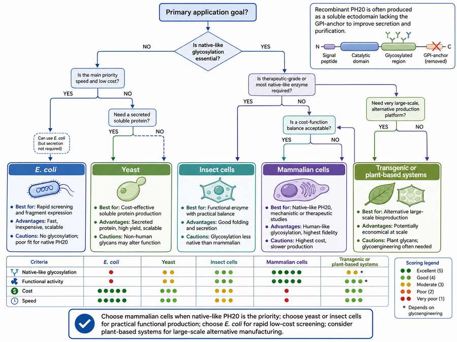
Fig 2. Application-based decision tree for selecting recombinant human hyaluronidase PH20
9. FAQ: Common Selection Questions
Q1: Can I use bovine testicular hyaluronidase instead of recombinant human PH20?
Bovine testicular hyaluronidase is a mixture of enzymes (including β-N-acetylhexosaminidase and β-glucuronidase) with broader substrate specificity and higher immunogenicity. While cost-effective for simple tissue dissociation, it is not recommended for mechanistic studies, cell-based assays, or in vivo applications where enzyme specificity and low endotoxin are critical.
Q2: Why does my hyaluronidase lose activity after storage?
Activity loss is most commonly caused by repeated freeze-thaw cycles, proteolytic degradation, or aggregation in low-ionic-strength buffers. Ensure aliquoting at first thaw, addition of glycerol (5–10%) or BSA (0.01%), and storage at −80°C for long-term retention.
Q3: How do I convert between USP units and TRUs?
There is no universal conversion factor because TRU definitions vary by substrate and protocol. The most reliable approach is to run both your sample and a USP-standardized hyaluronidase side-by-side under identical conditions and establish an empirical correlation curve.
Q4: Is glycosylation really necessary for PH20 activity?
Yes. N-glycosylation at specific asparagine residues is required for proper folding, secretion, and catalytic efficiency of PH20. Unglycosylated variants expressed in E. coli typically exhibit <10% of mammalian-expressed specific activity and are prone to aggregation.
Q5: What endotoxin level is safe for intravenous injection in mice?
For mouse studies, a conservative limit is <0.1 EU per dose (typically 10–50 μg protein). At higher endotoxin loads (>1 EU), mice may exhibit pyrogenic responses, cytokine storms, or lethality, confounding any interpretation of hyaluronidase pharmacology.
Q6: Should I worry about mycoplasma contamination?
While mycoplasma is primarily a concern for cell culture media, protein preparations derived from mammalian cells should be tested if the material will be used in long-term cell cultures or in vivo studies. PCR-based mycoplasma detection is rapid and sensitive.
Conclusion
Selecting the right recombinant human hyaluronidase requires balancing expression system fidelity, activity verification, purity, endotoxin control, and formulation stability against your specific experimental demands. Mammalian-derived PH20 (CHO or HEK) remains the gold standard for cell-based and in vivo research, while microbial systems offer cost-effective alternatives for biochemical screening. By applying the criteria outlined in this guide, researchers can minimize batch-to-batch variability, reduce assay artifacts, and generate reproducible, publication-quality data.