Human Hyaluronidase Family: Isoforms, Biological Functions, and Research Applications
A Comprehensive Review of HYAL Family Members, Tissue Distribution, and Biological Functions
Abstract
The human hyaluronidase (HYAL) family comprises six members—HYAL1, HYAL2, HYAL3, HYAL4, PH20/SPAM1, and HYALP1—that collectively regulate hyaluronan (HA) metabolism, extracellular matrix (ECM) remodeling, and numerous physiological processes. This review systematically examines the structural features, tissue distribution, enzymatic properties, and biological functions of each isoform. HYAL1 and HYAL2 serve as the principal somatic hyaluronidases responsible for HA catabolism, while PH20 exhibits unique neutral pH activity essential for fertilization. We discuss the roles of HYAL enzymes in tissue homeostasis, inflammation, wound repair, and pathological conditions including cancer and fibrosis. Additionally, we explore current research applications of recombinant human hyaluronidase in drug delivery and tissue permeability models, and identify critical open questions guiding future investigations.
human hyaluronidase family, HYAL1, HYAL2, PH20, hyaluronan metabolism, ECM remodeling, tumor microenvironment, drug delivery
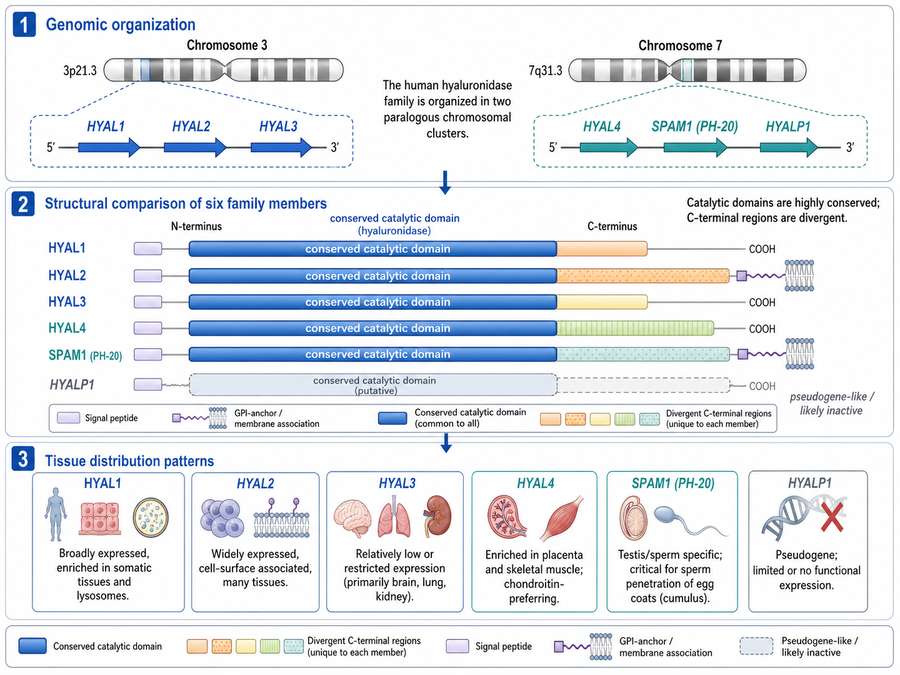
Fig 1. Genomic organization and structural features of the human hyaluronidase family. The six members are clustered on chromosomes 3p21.3 and 7q31.3, sharing conserved catalytic domains while differing in C-terminal regions and tissue distribution patterns.
1. Introduction: Overview of the Hyaluronidase Family
Hyaluronan (HA), a non-sulfated linear glycosaminoglycan composed of repeating disaccharide units of β-D-N-acetylglucosamine and D-glucuronic acid, represents a major component of the extracellular matrix (ECM) in vertebrate connective tissues. With a broad molecular weight range spanning from 10³ to 10⁷ Da, HA exhibits diverse physical and physiological properties that depend critically on its polymer length and local concentration.
The dynamic metabolism of HA is tightly controlled by three synthases (HAS1, HAS2, HAS3) and several hyaluronidases that mediate its catabolism. The human hyaluronidase family consists of six members encoded by genes clustered on chromosomes 3p21.3 (HYAL1, HYAL2, HYAL3) and 7q31.3 (HYAL4, PH20/SPAM1, HYALP1). These enzymes share approximately 33-44% sequence homology and belong to the glycosyl hydrolase family 56 (EC 3.2.1.35), functioning as endo-β-N-acetyl-hexosaminidases that hydrolyze the β1→4 glycosidic bonds of HA. While HYAL1 and HYAL2 are ubiquitously expressed and serve as the primary regulators of HA turnover in somatic tissues, other family members display restricted tissue distribution and specialized functions.
2. Human Hyaluronidase Isoforms
2.1 HYAL1
HYAL1 was the first human hyaluronidase to be characterized biochemically and remains the best-studied family member. The gene contains six exons producing a 2.6 kb mRNA that encodes a 436 amino acid protein. HYAL1 functions primarily as a lysosomal enzyme with optimal activity at acidic pH (3.5-3.8) and is widely distributed in plasma, urine, liver, kidney, spleen, and heart.
Enzymatically, HYAL1 degrades high molecular weight HA (HMW-HA) of any size to small oligosaccharides, predominantly tetrasaccharides (~0.8 kDa). This complete degradation capability makes HYAL1 the terminal enzyme in the HA catabolic pathway. Studies in HYAL1-deficient mice have revealed that loss of this enzyme results in osteoarthritis development and altered dendritic cell migration patterns, despite the animals being viable and fertile. A rare case of human HYAL1 deficiency demonstrated dramatically elevated plasma HA levels, confirming its essential role in systemic HA homeostasis.
2.2 HYAL2
HYAL2 contains four exons producing a 2 kb mRNA encoding a 473 amino acid protein that is anchored to the plasma membrane via a glycosylphosphatidylinositol (GPI) linkage. Based on mRNA expression patterns, HYAL2 represents the most widely expressed isoform in the body, detected in heart, skeletal muscle, colon, spleen, kidney, liver, placenta, and lungs.
Functionally, HYAL2 initiates HA catabolism by cleaving HMW-HA into intermediate-sized fragments of approximately 20 kDa (~50 disaccharide units). These fragments are subsequently internalized via receptor-mediated endocytosis (primarily through CD44) and delivered to lysosomes where HYAL1 completes their degradation to tetrasaccharides. HYAL2-deficient mice display significantly increased plasma HA levels, thickened endothelial glycocalyx, and severe cardiopulmonary dysfunction, underscoring its critical role in extracellular HA turnover.
2.3 HYAL3
HYAL3 is primarily distributed in testes and bone marrow, with weaker expression detected in various somatic tissues. Despite being an acid-active enzyme, HYAL3 exhibits no detectable in vitro hyaluronidase activity under standard assay conditions. Current evidence suggests that HYAL3 may function as a non-enzymatic regulator of HYAL1 activity rather than a direct HA-degrading enzyme. Notably, Hyal3-null mice do not accumulate HA and show only subtle changes in alveolar structure at advanced age, indicating that HYAL3 does not play a major role in constitutive HA degradation.
2.4 HYAL4
HYAL4 is predominantly expressed in skeletal muscle and placenta. Unlike other family members, HYAL4 does not cleave HA but instead functions as a chondroitinase with specific activity toward chondroitin sulfate (CS) and chondroitin sulfate proteoglycans. The enzyme contains a cysteine residue instead of the conserved Tyr247 found in other hyaluronidases, which likely contributes to its distinct substrate specificity. The biological significance of HYAL4 in CS metabolism remains an active area of investigation.
2.5 HYALP1 (PHYAL1)
HYALP1 represents an expressed pseudogene in humans, located on chromosome 7q31.3 alongside HYAL4 and PH20. Due to an exon deletion during human evolution, the amino acid translation process terminates prematurely, rendering the gene transcribed but not translated into functional protein. Interestingly, the murine ortholog of HYALP1 encodes an active enzyme in sperm, suggesting evolutionary divergence between species.
2.6 PH20 (SPAM1)
PH20, also known as Sperm Adhesion Molecule 1 (SPAM1), exhibits the strongest biological activity among all human hyaluronidases. It is predominantly expressed in testes and localized to the anterior head and acrosome of mature sperm. Unlike the acid-active HYAL1-3 enzymes, PH20 displays optimal activity at neutral pH (5.6-7.0) with a bimodal pH profile showing maxima at pH 4.5 and 7.5.
PH20 serves multiple functions during fertilization: it degrades the HA-rich cumulus oophorus surrounding the oocyte, enabling sperm penetration, and subsequently acts as a receptor for sperm binding to the zona pellucida. Beyond reproduction, PH20 expression has been detected in demyelinating lesions where elevated levels inhibit oligodendrocyte progenitor cell maturation and remyelination.
| Isoform | Chromosomal Location | Primary Tissues | Optimal pH | Substrate/Product | Membrane Association |
|---|---|---|---|---|---|
| HYAL1 | 3p21.3 | Liver, kidney, spleen, plasma, urine | 3.5-3.8 | HA → Tetrasaccharides | Soluble/Lysosomal |
| HYAL2 | 3p21.3 | Heart, muscle, kidney, liver, lung, spleen | <4.0-6.0 | HMW-HA → ~20 kDa fragments | GPI-anchored |
| HYAL3 | 3p21.3 | Testes, bone marrow, brain (low) | Acidic | No detectable HA activity | Unknown |
| HYAL4 | 7q31.3 | Skeletal muscle, placenta | Acidic | Chondroitin sulfate | Unknown |
| PH20 | 7q31.3 | Testes, sperm | 4.5 & 7.5 (bimodal) | HA → Various oligosaccharides | GPI-anchored |
| HYALP1 | 7q31.3 | Transcribed only (pseudogene) | N/A | Non-functional | N/A |
3. Hyaluronan Metabolism and ECM Remodeling
HA catabolism represents a highly coordinated two-stage process involving sequential enzymatic activities and cellular trafficking mechanisms. In the extracellular matrix, HMW-HA (typically >1000 kDa) provides structural integrity, tissue hydration, and anti-inflammatory signaling through interactions with CD44 and RHAMM receptors.
The catabolic cascade begins with membrane-anchored HYAL2 cleaving HMW-HA into ~20 kDa fragments at the cell surface. These intermediate fragments, bound to CD44, undergo receptor-mediated endocytosis and are transported to late endosomes and lysosomes. Within the acidic lysosomal environment (pH 4.5-5.0), HYAL1 completes the degradation process, generating biologically active tetrasaccharides that can be further processed by exoglycosidases (β-D-glucuronidase and β-N-acetyl-hexosaminidase) into monosaccharides.
The size of HA fragments critically determines their biological activity. High molecular weight HA (>500 kDa) typically exhibits anti-inflammatory, anti-angiogenic, and immunosuppressive properties, whereas low molecular weight fragments (20-200 kDa) generated by hyaluronidase activity promote inflammation, angiogenesis, and cellular proliferation.
Beyond the classical HYAL pathway, two novel HA-degrading enzymes have been identified: CEMIP (also called KIAA1199 or HYBID) and TMEM2. CEMIP, a 150 kDa secreted protein with no sequence homology to HYAL family members, mediates HA depolymerization via clathrin-coated pit endocytosis. TMEM2, a type II transmembrane protein, cleaves HMW-HA into ~5 kDa fragments in a Ca²⁺-dependent manner under neutral pH conditions. These alternative pathways may be particularly relevant in specific tissues or pathological contexts where HYAL activity is compromised.
4. PH20 and Other HYAL Members: Key Differences
PH20 stands apart from somatic hyaluronidases in several critical aspects. While HYAL1 and HYAL2 require acidic environments for optimal catalytic activity, PH20 maintains robust enzymatic function at neutral pH, a property essential for its role in fertilization within the female reproductive tract. Structurally, PH20 contains two additional cysteine residues in its C-terminal region that do not align with other family members, potentially contributing to its unique membrane topology and substrate interactions.
Enzymatically, bovine PH20 (which shares 61% sequence identity with human PH20) demonstrates approximately 400-fold greater activity than human HYAL2 under acidic conditions, highlighting the exceptional catalytic efficiency of this isoform. Unlike the somatic hyaluronidases involved in routine HA turnover, PH20 expression is essentially restricted to male germ cells, although ectopic expression has been reported in certain pathological conditions.
| Feature | HYAL1/HYAL2 | PH20 |
|---|---|---|
| Primary Function | Somatic HA catabolism | Fertilization (cumulus penetration) |
| pH Optimum | Acidic (3.5-4.0) | Neutral (5.6-7.5) |
| Tissue Distribution | Ubiquitous (most tissues) | Testes-specific |
| Cellular Location | Lysosomal (HYAL1) / Cell surface (HYAL2) | Acrosomal membrane |
| Catalytic Efficiency | Moderate | Very high (~400× HYAL2) |
| Clinical Application | Cancer biomarkers, lysosomal disorders | Assisted reproduction, drug dispersion |
5. Physiological Functions
5.1 Fertilization
PH20 plays an indispensable role in mammalian fertilization through its dual functionality. During sperm transit through the female reproductive tract, PH20 remains inactive due to membrane compartmentalization. Upon capacitation and the acrosome reaction, PH20 redistributes to the anterior sperm head surface where it degrades the HA-rich cumulus oophorus, creating a path for sperm penetration toward the oocyte. Following cumulus penetration, PH20 mediates sperm-zona pellucida binding through its HA-binding domain, functioning as a signaling receptor that triggers downstream events essential for gamete fusion.
5.2 Tissue Remodeling
HA metabolism is fundamental to tissue morphogenesis and remodeling during embryonic development and adult tissue maintenance. HYAL1 and HYAL2 coordinate the precise temporal and spatial degradation of HA that is necessary for cell migration, tissue patterning, and organogenesis. The half-life of HA varies dramatically between tissues—from 2-3 hours in epidermis to approximately 1 day in dermis—reflecting tissue-specific rates of synthesis and catabolism.
5.3 Inflammation
Hyaluronidases actively participate in inflammatory responses through the generation of bioactive HA fragments. During acute inflammation, pro-inflammatory cytokines (TNF-α, IL-1β) activate NF-κB signaling pathways, leading to upregulated expression of HYAL1, HYAL2, and HYAL3. The resulting LMW-HA fragments function as damage-associated molecular patterns (DAMPs), stimulating monocyte activation through Toll-like receptor 4 (TLR4) and promoting endothelial cell proliferation and migration. Conversely, during inflammation resolution, anti-inflammatory cytokines such as IL-10 suppress hyaluronidase production, restoring HMW-HA homeostasis.
5.4 Wound Repair
Following tissue injury, coordinated HA synthesis and degradation orchestrate the sequential phases of wound healing. Initial inflammation triggers HA accumulation that facilitates leukocyte infiltration and pathogen clearance. Subsequent hyaluronidase-mediated degradation generates LMW-HA fragments that stimulate fibroblast migration, angiogenesis, and granulation tissue formation. In the final remodeling phase, restored HYAL activity ensures removal of excess HA and transition to scar tissue. Dysregulated hyaluronidase activity during wound healing can result in chronic wounds or excessive scarring.
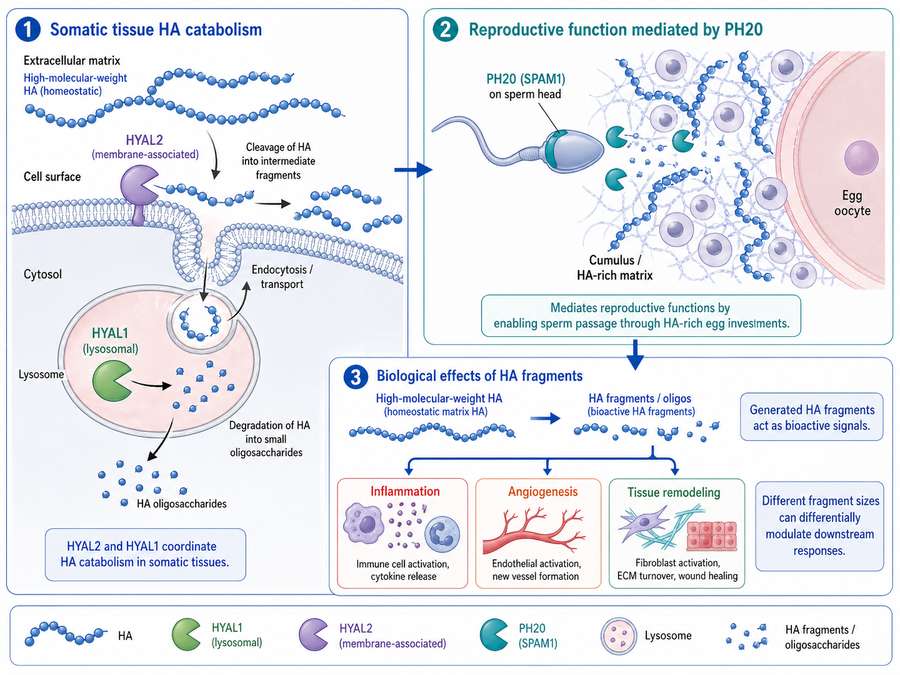
Fig 2. Overview of physiological processes regulated by the human hyaluronidase family. HYAL1 and HYAL2 coordinate HA catabolism in somatic tissues, while PH20 mediates reproductive functions. Generated HA fragments differentially modulate inflammation, angiogenesis, and tissue remodeling.
6. Pathological Relevance
6.1 Tumor Microenvironment
Altered hyaluronidase expression and activity significantly contribute to cancer progression and metastasis. HYAL1 is frequently overexpressed in prostate, bladder, and head and neck cancers, where elevated serum levels correlate with tumor grade and poor prognosis. Paradoxically, HYAL1 and HYAL2 can also function as tumor suppressors in certain contexts, as both genes are located in the 3p21.3 LUCA region—a chromosomal area commonly deleted in lung and other cancers. This dichotomy reflects the complex role of HA metabolism in cancer, where the balance between HMW-HA (anti-tumorigenic) and LMW-HA (pro-tumorigenic) fragments determines disease outcome.
HYAL2 additionally serves as the cellular receptor for Jaagsiekte sheep retrovirus (JSRV), and its sequestration by viral envelope proteins leads to ligand-independent activation of the RON receptor tyrosine kinase and downstream AKT/MAPK signaling pathways that promote cellular transformation.
6.2 Fibrosis
Dysregulated HA accumulation and impaired hyaluronidase activity characterize multiple fibrotic diseases. In pulmonary fibrosis, altered HYAL1/HYAL2 expression disrupts normal HA turnover, leading to excessive ECM deposition and progressive tissue scarring. Similarly, liver fibrosis involves disturbed HA metabolism where elevated serum HA levels serve as clinical biomarkers of disease severity. Understanding the precise mechanisms by which hyaluronidases regulate fibrotic processes may reveal novel therapeutic targets.
6.3 Inflammatory Diseases
Aberrant hyaluronidase activity contributes to the pathogenesis of various inflammatory conditions. In osteoarthritis, HYAL1 deficiency leads to HA accumulation in articular cartilage, accelerating joint degeneration. In inflammatory skin disorders, keratinocyte-derived HYAL1 and HYAL2 regulate epidermal HA content, with imbalances contributing to barrier dysfunction. The pro-inflammatory LMW-HA fragments generated by excessive hyaluronidase activity can perpetuate chronic inflammation in diseases such as rheumatoid arthritis and inflammatory bowel disease.
| Disease | Involved Isoform(s) | Pathological Mechanism | Clinical Relevance |
|---|---|---|---|
| Prostate Cancer | HYAL1 | Overexpression generates pro-angiogenic HA fragments | Serum biomarker for tumor grade |
| Lung Cancer | HYAL1, HYAL2 | 3p21.3 deletion inactivates tumor suppressor function | Prognostic indicator |
| Osteoarthritis | HYAL1 | Deficiency causes HA accumulation in cartilage | Therapeutic target |
| Pulmonary Fibrosis | HYAL1, HYAL2 | Impaired HA turnover promotes ECM deposition | Biomarker development |
| Multiple Sclerosis | PH20 | Ectopic expression inhibits remyelination | Novel therapeutic avenue |
| Lysosomal Storage Disorders | HYAL1 | Deficiency leads to HA accumulation in tissues | Enzyme replacement therapy |
7. Research Applications
7.1 Substrate Degradation Studies
Recombinant human hyaluronidase preparations serve as essential tools for investigating HA structure-function relationships in vitro. The ability to specifically degrade HMW-HA to defined fragment sizes enables researchers to dissect the differential signaling properties of various HA polymers. Such studies have been instrumental in establishing that LMW-HA fragments promote cell migration and proliferation, while HMW-HA maintains quiescent phenotypes.
7.2 Tissue Permeability Models
Hyaluronidases are widely employed to enhance tissue permeability in experimental settings. By degrading HA in the ECM interstitial matrix, these enzymes increase the effective pore size of tissue barriers, facilitating the diffusion of macromolecules, nanoparticles, and diagnostic agents. This application is particularly valuable in tumor penetration studies, where the dense HA-rich stroma presents a significant barrier to drug delivery.
7.3 Drug Delivery Research
The clinical utility of hyaluronidase as a spreading factor has been recognized for decades, and recombinant human hyaluronidase is now employed as an adjuvant to increase the absorption and dispersion of co-injected drugs. Current research focuses on developing hyaluronidase-triggered drug release systems, where HA-based nanoparticles remain stable in circulation but release therapeutic payloads upon encountering tumor-associated hyaluronidases. Additionally, hyaluronidase pretreatment of solid tumors is being explored as a strategy to normalize the tumor microenvironment and improve the efficacy of chemotherapy and immunotherapy agents.
Recombinant human PH20 (rHuPH20) is clinically approved as an adjuvant for subcutaneous drug administration, where it temporarily depolymerizes subcutaneous HA to increase the dispersion and absorption of co-formulated biologics. This technology has been successfully applied to subcutaneous formulations of trastuzumab, rituximab, and immunoglobulins.
8. Open Questions and Future Research Directions
Despite significant advances in understanding the human hyaluronidase family, several critical questions remain unanswered:
- HYAL3 Function: The precise physiological role of HYAL3 remains enigmatic. Does it function as a regulatory protein modulating HYAL1 activity, or does it possess cryptic enzymatic activity under specific conditions yet to be identified?
- Tissue-Specific Regulation: How do different tissues coordinate the expression and activity of multiple hyaluronidase isoforms to achieve precise control of HA metabolism? What transcriptional and post-translational mechanisms govern this regulation?
- Fragment Signaling: What are the specific receptors and signaling pathways differentially activated by distinct HA oligosaccharide sizes generated through hyaluronidase activity?
- Therapeutic Targeting: Can isoform-specific hyaluronidase inhibitors be developed for conditions characterized by excessive HA degradation (e.g., cancer, inflammation) without disrupting essential physiological HA turnover?
- Non-Enzymatic Functions: Do hyaluronidases possess signaling or scaffolding functions independent of their catalytic activity, particularly given the conserved C-terminal EGF-like domains?
- CEMIP/TMEM2 Integration: How do the novel HA-degrading enzymes CEMIP and TMEM2 functionally interact with the classical HYAL pathway in tissue-specific contexts?
Future research addressing these questions will require integrative approaches combining structural biology, genetically modified animal models, and advanced analytical techniques for HA fragment characterization. The development of isoform-specific activity assays and selective inhibitors will be particularly crucial for dissecting the individual contributions of HYAL family members in health and disease.
Conclusion
The human hyaluronidase family represents a critical enzymatic network governing hyaluronan metabolism, extracellular matrix dynamics, and numerous physiological processes. From the ubiquitous somatic enzymes HYAL1 and HYAL2 to the specialized reproductive enzyme PH20, each family member contributes uniquely to HA homeostasis. Understanding the distinct biochemical properties, tissue distributions, and biological functions of these enzymes provides essential insights into both normal physiology and disease pathogenesis. As research tools, recombinant human hyaluronidase preparations continue to enable breakthrough discoveries in drug delivery, tissue engineering, and cancer biology. Addressing the open questions in hyaluronidase biology will undoubtedly yield novel therapeutic strategies for diseases ranging from cancer and fibrosis to inflammatory disorders and reproductive medicine.
For precision medicine research, understanding the isoform-specific functions of the hyaluronidase family is essential for developing targeted therapeutic interventions. We encourage researchers to leverage high-quality recombinant hyaluronidase products to advance mechanistic studies and translational applications in hyaluronan biology.
References
1. Stern, R., et al. (2007). The hyaluronidases: Their genomics, structures, and mechanisms of action. Chemical Reviews, 107(7), 2222-2245.
2. Csoka, A. B., Scherer, S. W., & Stern, R. (1999). Expression analysis of six paralogous human hyaluronidase genes clustered on chromosomes 3p21 and 7q31. Genomics, 60(3), 356-361.
3. Hyaluronidase: structure, mechanism of action, diseases and therapeutic targets. PMC, PMC12254123.
4. Hyaluronan: Metabolism and Function. PMC - NIH, PMC7695009.
5. Structure of Human Hyaluronidase-1. ACS Publications, Biochemistry, 2007.
6. HYAL1 and HYAL2 Inhibit Tumour Growth In Vivo but Not In Vitro. PMC, PMC2516603.
7. The Hyaluronidases: Their Genomics, Structures, and Mechanisms. PMC, PMC2547145.
8. Antimicrobial Activity Versus Virulence Potential of Hyaluronic Acid. MDPI International Journal of Molecular Sciences, 2025.
9. The Degradation of Hyaluronan in the Skin. MDPI Biomolecules, 2022.
10. Subtype Specific Elevated Expression of Hyaluronidase-1 in Epithelial Ovarian Cancer. PMC, PMC3112150.
11. Leiden University Repository. Hyaluronan metabolism and endothelial function.
12. Polish Journal of Pharmacology. Hyaluronan catabolism and hyaluronidases.
13. University of Liverpool Repository. HYAL family in cancer progression.
14. University of Ferrara Thesis. HYAL roles in HA metabolism.
15. University of Eastern Finland Repository. Hyaluronan degradation mechanisms.
16. Epidermal Hyaluronan in Barrier Alteration-Related Disease. MDPI Cells, 2021.
17. ScienceDirect Topics. Hyaluronan-mediated motility receptor overview.