Troubleshooting Recombinant Human Hyaluronidase Experiments
Low Activity, High Background, and Variability
Overview: Most Common Hyaluronidase Assay Problems
Recombinant human hyaluronidase is widely used in hyaluronic acid (HA) degradation assays, drug dispersion studies, and tissue permeability research. However, researchers frequently encounter three categories of assay failure: low or no detectable enzyme activity, elevated background signal, and poor replicate consistency. This troubleshooting guide systematically addresses each failure mode with actionable diagnostic steps and corrective measures.
Over 60% of hyaluronidase troubleshooting cases are resolved by correcting substrate preparation, buffer pH, or enzyme storage conditions—not by replacing the enzyme itself.
Table 1. Frequency of Reported Hyaluronidase Assay Failures
| Failure Category | Reported Frequency (%) | Typical Root Cause | Resolution Rate (%) |
|---|---|---|---|
| Low or no enzyme activity | 38 | Enzyme inactivation or incorrect pH | 85 |
| High background signal | 27 | Substrate impurities or detection interference | 78 |
| Poor replicate consistency | 21 | Pipetting error or uneven mixing | 92 |
| Substrate-related anomalies | 9 | HA molecular weight variability | 70 |
| Temperature or storage issues | 5 | Freeze-thaw damage or thermal drift | 88 |
1. Low or No Enzyme Activity: Causes and Diagnostic Steps
1.1 Enzyme Integrity and Storage Verification
The most common cause of low hyaluronidase activity is protein denaturation due to improper handling. Human recombinant hyaluronidase (PH20) is a glycoprotein sensitive to repeated freeze-thaw cycles and prolonged exposure to room temperature.
- Check aliquot history: If the vial has undergone more than two freeze-thaw cycles, activity loss of 15–40% is expected. Always prepare single-use aliquots upon first thaw.
- Verify storage temperature: Lyophilized enzyme should be stored at −20°C or below. Reconstituted enzyme in stabilizing buffer (e.g., 20 mM sodium phosphate, pH 7.4, with 0.1% BSA) should be used within 24 hours or flash-frozen in liquid nitrogen.
- Inspect for precipitation: Visible aggregates or turbidity indicate irreversible denaturation. Centrifuge at 10,000 × g for 5 min and assay the supernatant to confirm.
1.2 Buffer and pH Compatibility
Human hyaluronidase PH20 exhibits a narrow pH optimum between 4.5 and 6.0, with maximal activity at approximately pH 5.0. Assays conducted at neutral or alkaline pH (pH 7.0–8.0) can show >80% activity loss.
- Confirm assay buffer pH: Use a calibrated pH meter immediately before the reaction. Acetate or citrate buffers (50 mM, pH 5.0) are recommended. Phosphate buffers at pH 7.4 are suitable only for stock enzyme dilution, not for the catalytic reaction.
- Check ionic strength: High salt concentrations (>200 mM NaCl) can inhibit hyaluronidase by stabilizing the HA polymer structure. Maintain ionic strength between 50 and 150 mM.
- Avoid chelating agents: EDTA or EGTA at concentrations >1 mM can sequester trace metal ions (Ca²⁺, Mg²⁺) that may serve as cofactors for optimal enzyme conformation.
1.3 Substrate Concentration and Quality
HA substrate must be present at saturating concentrations. Below the Km (~0.5–1.0 mg/mL for high-molecular-weight HA), the reaction rate becomes substrate-limited and may appear falsely low.
- Verify HA concentration: Use a validated stock solution at 2–5 mg/mL. HA is hygroscopic; weigh accurately and dissolve in assay buffer with gentle rotation overnight at 4°C.
- Check substrate lot: Switch to a fresh HA preparation if the current lot is >6 months old or has been stored without desiccant.
If activity is zero across all samples, test a positive control (commercial hyaluronidase reference standard) in parallel. If the control is also inactive, the problem lies in the assay buffer, substrate, or detection system—not your recombinant human hyaluronidase sample.
2. High Background Signal: Substrate, Buffer, and Detection System Issues
2.1 Substrate Purity and Autodegradation
Commercial HA preparations may contain low-molecular-weight fragments or endotoxins that generate signal in the absence of enzyme. This is especially problematic in turbidimetric or colorimetric endpoint assays.
- Run a substrate-only blank: Include a well containing HA substrate and buffer without enzyme. If absorbance or turbidity is elevated (>10% of expected signal), the substrate is contaminated or partially degraded.
- Filter substrate stock: Pass HA solution through a 0.22 μm filter to remove particulates. Avoid excessive pressure to prevent shear-induced fragmentation.
- Check HA source: Bacterial fermentation-derived HA (Streptococcus) may contain trace protein impurities. Animal-derived HA (rooster comb) carries higher endotoxin risk. Highly purified recombinant HA is preferred for sensitive assays.
2.2 Detection System Interference
Common detection methods for hyaluronidase activity include the Morgan–Elson colorimetric assay (measuring N-acetylglucosamine release), turbidimetry with cetylpyridinium chloride (CPC) precipitation, and ELISA-based HA competition assays.
- Morgan–Elson assay: Background can arise from non-enzymatic release of reducing sugars during boiling. Ensure the pH of the borate buffer is exactly 9.1–9.2; deviations cause premature color development.
- CPC turbidimetry: Unreacted HA forms a turbid complex with CPC. High background indicates insufficient reaction time or enzyme concentration that is too low. Conversely, over-digestion can also cause low signal if all HA is degraded below the CPC-binding threshold.
- ELISA interference: Serum components or BSA in the enzyme diluent can cross-react with anti-HA antibodies. Use a matched diluent without carrier protein, or include an isotype-matched irrelevant antibody control.
Table 2. Background Signal Sources by Detection Method
| Detection Method | Typical Background Source | Corrective Action |
|---|---|---|
| Morgan–Elson (colorimetric) | Non-enzymatic sugar release at high pH | Strict pH control; include buffer-only blank |
| CPC turbidimetry | Particulate HA or premature precipitation | Filter substrate; standardize mixing speed |
| ELISA (competitive) | Antibody cross-reactivity with diluents | Use protein-free diluent; add blocking step |
| Fluorometric (HA-fluorescein) | Auto-fluorescence of buffer additives | Switch to low-fluorescence buffer; read blank |
3. Poor Replicate Consistency: Pipetting, Mixing, and Timing Control
3.1 Pipetting Accuracy and Technique
Human hyaluronidase assay variability often originates at the pipetting stage. HA solutions are viscous (especially at >2 mg/mL), and small volume errors propagate nonlinearly into large signal deviations.
- Use positive-displacement pipettes: For viscous HA stocks, air-displacement pipettes can deliver 5–15% less volume than set. Positive-displacement tips (e.g., Microman, Gilson) or reverse-pipetting technique is strongly recommended.
- Pre-wet tips: Aspirate and dispense HA solution once before the final transfer to coat the tip interior and reduce adhesion.
- Calibrate pipettes quarterly: Even slight drift in piston seals can cause systematic bias across replicates.
3.2 Mixing and Reaction Uniformity
Incomplete mixing creates localized concentration gradients, leading to variable reaction kinetics.
- Mix by gentle pipetting: After adding enzyme to substrate, pipette up and down 3–5 times with a volume equal to 50% of the total reaction volume. Avoid vortexing, which can denature the enzyme through shear stress and air–liquid interface exposure.
- Staggered timing: When running multiple plates or time points, use a multichannel pipette or automated liquid handler to minimize well-to-well timing differences. A 30-second stagger across a 96-well plate can cause 10% CV in endpoint assays.
3.3 Incubation Time and Temperature Uniformity
Hyaluronidase activity is temperature-sensitive, with a Q10 of approximately 2.0 between 25°C and 37°C. A 1°C deviation can alter reaction rate by 7–10%.
- Use a calibrated incubator or water bath: Verify temperature with a NIST-traceable thermometer placed at the plate level, not just the ambient air sensor.
- Avoid plate-edge effects: Peripheral wells in microplates may be 1–2°C cooler due to evaporative cooling. Include edge wells as blanks or fill them with buffer to insulate interior assay wells.
- Quench simultaneously: For endpoint assays, add stop solution (e.g., 0.2 M NaOH or boiling) to all wells at exactly the same elapsed time using a multichannel pipette or reagent dispenser.
4. Substrate-Related Problems: HA Molecular Weight, Solubility, and Lot-to-Lot Variability
4.1 HA Molecular Weight Selection
Hyaluronidase cleaves the β-1,4-glycosidic bond between N-acetylglucosamine and glucuronic acid. The rate of cleavage depends on HA chain length: high-molecular-weight (HMW) HA (>1,000 kDa) is degraded more slowly than low-molecular-weight (LMW) HA (<100 kDa) due to steric hindrance and reduced enzyme accessibility.
- Match HA MW to assay sensitivity: For kinetic assays measuring initial velocity, use LMW HA (100–300 kDa) to ensure linear progress curves within 30–60 minutes. For endpoint assays requiring robust signal, HMW HA (1,000–2,000 kDa) provides larger turbidity changes but requires longer digestion.
- Document HA specifications: Record the nominal MW, polydispersity index (PDI), and source for every lot. Do not mix lots within the same experiment.
4.2 Solubility and Preparation Artifacts
HA is notoriously difficult to dissolve. Rapid addition of powder to aqueous buffer creates clumps that hydrate externally but remain dry internally, leading to heterogeneous substrate distribution.
- Dissolve by slow hydration: Sprinkle HA powder onto the surface of cold buffer (4°C) and allow passive hydration for 2–4 hours with gentle rocking. Do not stir vigorously or vortex.
- Check for undissolved particles: Inspect the solution against a dark background. Particulates will cause localized high-substrate zones and erratic enzyme activity.
Table 3. HA Molecular Weight vs. Recommended Assay Parameters
| HA Molecular Weight | Optimal Enzyme Conc. | Recommended Incubation | Best Detection Method |
|---|---|---|---|
| Low (50–100 kDa) | 10–50 U/mL | 15–30 min at 37°C | Morgan–Elson (sensitive) |
| Medium (300–800 kDa) | 50–200 U/mL | 30–60 min at 37°C | CPC turbidimetry |
| High (>1,000 kDa) | 200–500 U/mL | 60–120 min at 37°C | CPC turbidimetry or viscometry |
5. Temperature and pH Effects on Hyaluronidase Activity
5.1 Temperature Optimum and Thermal Stability
Recombinant human PH20 displays maximal activity at 37°C, with 50% activity at 25°C and near-complete inactivation above 50°C. However, prolonged incubation at 37°C (>4 hours) can lead to gradual activity loss due to thermal denaturation.
- Optimize reaction duration: For standard assays, limit incubation to 30–90 minutes. For extended digestions, supplement with 0.01% BSA or 5% glycerol as thermal stabilizers.
- Avoid temperature fluctuations: Do not remove plates from the incubator for intermediate readings unless the assay is designed for kinetic monitoring at room temperature.
5.2 pH Sensitivity and Buffer Selection
The pH profile of human hyaluronidase is bell-shaped, with rapid decline below pH 4.0 and above pH 7.0. Buffer selection must consider both pH capacity and ionic composition.
- Acetate buffer (50 mM, pH 5.0): Recommended for most turbidimetric and colorimetric assays. Avoid acetate in Morgan–Elson assays because acetate ions interfere with the borate-catalyzed color reaction.
- Citrate–phosphate buffer (McIlvaine's, pH 5.0–6.0): Excellent buffering capacity but may chelate metal ions. Use only if metal cofactors are not required.
- HEPES or MES buffers: Acceptable at pH 5.5–6.5, though not optimal. Use only if acetate is incompatible with downstream detection chemistry.
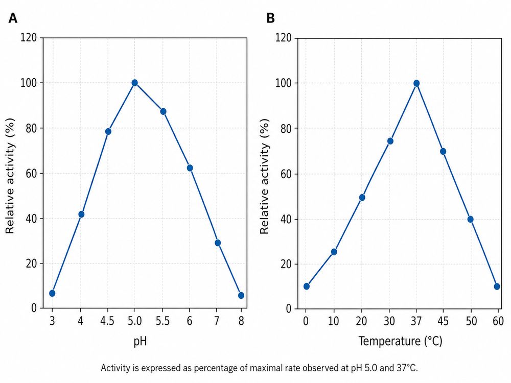
Fig 1. Effect of pH and temperature on recombinant human hyaluronidase (PH20) activity. Activity is expressed as percentage of maximal rate observed at pH 5.0 and 37°C.
6. Storage and Freeze-Thaw Effects on Enzyme Stability
6.1 Lyophilized vs. Liquid Formulations
Lyophilized recombinant human hyaluronidase is significantly more stable than liquid formulations. When stored at −20°C in a desiccated environment, lyophilized enzyme retains >95% activity for 24 months. In contrast, liquid enzyme in aqueous buffer loses 10–20% activity per month at 4°C and 40–60% per freeze-thaw cycle at −20°C.
- Reconstitution protocol: Reconstitute lyophilized enzyme with sterile, chilled buffer (4°C). Allow 10–15 minutes for complete dissolution without agitation. Do not vortex or shake vigorously.
- Aliquot immediately: Divide reconstituted enzyme into 10–20 μL single-use aliquots in PCR tubes or low-protein-binding microcentrifuge tubes. Flash-freeze in liquid nitrogen and store at −80°C.
6.2 Freeze-Thaw Damage Mechanisms
Ice crystal formation during slow freezing ruptures protein hydration shells and promotes intermolecular aggregation. PH20 is particularly susceptible because its glycosylation pattern influences solubility and conformational stability.
- Use cryoprotectants: Add 10–20% glycerol or 0.5% BSA to the storage buffer if multiple freeze-thaw cycles are unavoidable. These additives stabilize the protein against ice–water interface denaturation.
- Thaw on ice: Never thaw enzyme at 37°C or room temperature. Place aliquots on wet ice and allow 15–20 minutes for gradual thawing. Use immediately; do not refreeze.
Table 4. Storage Condition Impact on Hyaluronidase Activity Retention
| Storage Condition | 1 Month | 3 Months | 6 Months | 12 Months |
|---|---|---|---|---|
| Lyophilized, −20°C, desiccated | 98% | 97% | 96% | 95% |
| Liquid, −80°C, 10% glycerol | 96% | 94% | 91% | 88% |
| Liquid, −20°C, no cryoprotectant | 85% | 62% | 40% | 15% |
| Liquid, 4°C | 90% | 72% | 50% | 25% |
| Repeated freeze-thaw (−20°C, no protectant) | 60% (after 1 cycle) | 35% (after 3 cycles) | 10% (after 5 cycles) | <5% (after 7 cycles) |
7. Control Design Problems
7.1 Essential Controls for Every Assay
A robust hyaluronidase assay requires three categories of controls: positive, negative, and internal reference. Omitting any category invalidates data interpretation and complicates troubleshooting.
- Positive control: A characterized reference hyaluronidase (e.g., bovine testicular hyaluronidase or a validated recombinant PH20 standard) run at a fixed concentration. This confirms that the substrate, buffer, and detection system are functional.
- Negative control (enzyme blank): Assay buffer + substrate, no enzyme. This defines the baseline signal and detects substrate autodegradation or contamination.
- Buffer blank: Assay buffer only, no substrate or enzyme. This corrects for instrument drift, plate absorbance, or reagent color.
- Internal reference: A mid-range enzyme concentration included on every plate to monitor inter-plate and inter-day variability. Target CV < 10% for this reference across all runs.
7.2 Common Control Failures
- Positive control is low, test sample is low: Problem is systemic (buffer, substrate, detection, or temperature). Do not blame the test enzyme.
- Positive control is normal, test sample is low: Problem is specific to the test enzyme (storage damage, concentration error, or inhibitor contamination).
- Negative control is high: Substrate is degraded or contaminated; detection reagents are faulty; or the stop/quench step failed.
- High variability in internal reference: Pipetting inconsistency, uneven incubation temperature, or plate-position effects.
8. Troubleshooting Decision Tree
Use the following decision framework to narrow down the root cause systematically:
Step 1: Is the positive control active?
NO → The problem is systemic. Proceed to Step 2.
YES → The problem is specific to your test sample. Proceed to Step 5.
Step 2: Is the negative control (substrate + buffer) elevated?
YES → Substrate is degraded or contaminated. Replace HA lot; filter stock; check storage.
NO → Substrate is acceptable. Proceed to Step 3.
Step 3: Is the buffer blank elevated?
YES → Detection reagents are faulty or contaminated. Prepare fresh reagents; check instrument calibration.
NO → Detection system is clean. Proceed to Step 4.
Step 4: Check pH, temperature, and timing
Verify assay buffer pH with a calibrated meter (target pH 5.0 ± 0.2).
Verify incubator temperature with a NIST-traceable thermometer (target 37°C ± 0.5°C).
Verify reaction time with a stopwatch (staggered additions require correction).
If all parameters are correct, replace the enzyme with a fresh aliquot.
Step 5: Test sample-specific diagnostics
Check enzyme storage history (freeze-thaw count, temperature).
Visually inspect for precipitation or turbidity.
Re-quantify enzyme concentration (Bradford, BCA, or A280).
Test at 2× and 0.5× nominal concentration to rule out inhibition or substrate depletion.
If all checks pass, request a replacement vial from the supplier.
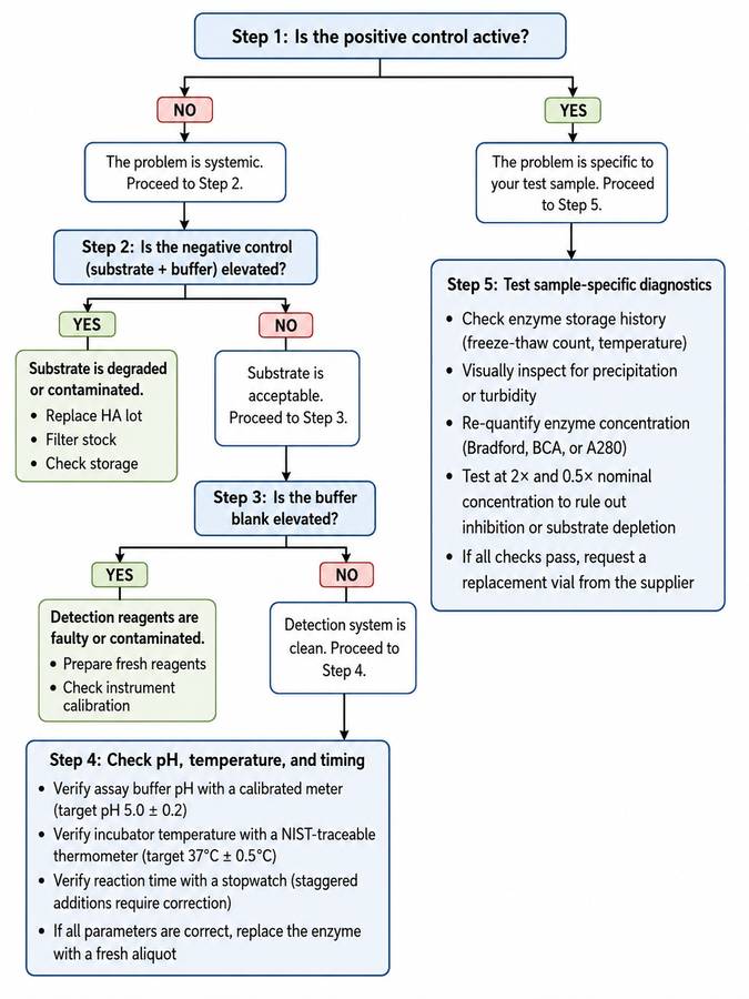
Fig 2. Troubleshooting decision tree for hyaluronidase assay failures. Start at the top and follow the branches based on your control results.
9. Preventive Checklist Before Running the Next Assay
Complete this checklist before initiating any hyaluronidase assay to prevent the most common sources of failure:
Enzyme Preparation
- Enzyme is from a fresh aliquot with ≤1 freeze-thaw cycle
- Enzyme concentration verified by A280 or protein assay
- No visible precipitation, turbidity, or color change in enzyme solution
- Enzyme diluted in recommended assay buffer immediately before use
Substrate Preparation
- HA stock dissolved by passive hydration at 4°C for ≥2 hours
- HA concentration confirmed by uronic acid assay or validated stock label
- No undissolved particles or fibers visible in substrate solution
- Substrate lot documented; same lot used for entire experiment
Buffer and Reagents
- Assay buffer pH measured and confirmed at assay temperature (37°C)
- Buffer ionic strength between 50–150 mM; no chelating agents >1 mM
- Detection reagents freshly prepared or validated for stability
- All reagents equilibrated to room temperature before pipetting (except enzyme)
Equipment and Environment
- Pipettes calibrated within the last 3 months; positive-displacement tips available for viscous solutions
- Incubator or water bath temperature verified at plate level (37°C ± 0.5°C)
- Microplate reader baseline checked with buffer blank; no well-to-well optical artifacts
- Timer or automated liquid handler programmed for synchronized reagent addition and quenching
Controls and Documentation
- Positive control (reference hyaluronidase) included on every plate
- Negative control (substrate + buffer, no enzyme) included in triplicate
- Buffer blank included for baseline subtraction
- Internal reference at mid-range concentration for inter-run tracking
- All lot numbers, pipette settings, incubation times, and temperatures recorded in lab notebook or ELN
When in doubt, run a mini-pilot with your positive control, negative control, and one test sample before committing the full plate. A 30-minute pilot can save hours of failed data and wasted recombinant human hyaluronidase reagent.
References
1. Frost, G. I., et al. (2013). Adv Drug Deliv Rev, 65(13-14): 1861-1867.
2. Bookbinder, L. H., et al. (2006). J Biol Chem, 281(48): 36589-36599.
3. Stern, R. (2014). Matrix Biol, 35: 9-12.
4. European Pharmacopoeia 11.0, Hyaluronidase monograph.
5. Chang, D. S., et al. (2019). Anal Biochem, 582: 113357.
6. Tiwari, S., et al. (2020). J Pharm Biomed Anal, 186: 113286.