
Structural Features of Acid Alpha-Glucosidase and Their Impact on Enzyme Function
How architecture, glycosylation, folding, and active-site organization shape lysosomal GAA performance
Abstract
Acid alpha-glucosidase (GAA) is the lysosomal hydrolase responsible for converting glycogen-derived oligosaccharides into free glucose under acidic conditions. Because GAA function depends on correct folding, proteolytic maturation, glycosylation, mannose-6-phosphate (M6P) tagging, and the integrity of its catalytic pocket, structure is inseparable from biology. For researchers working with recombinant human GAA, understanding these structural features helps explain why apparently similar enzyme preparations can differ in uptake, intracellular delivery, activity, and stability.
This technical article summarizes the domain organization of human GAA, distinguishes precursor and mature lysosomal forms, explains how glycosylation and M6P support trafficking, and connects active-site geometry to glycogen hydrolysis. It also highlights how structural instability, misfolding, or altered post-translational modification can reduce function and how these insights guide research use of acid alpha-glucosidase and next-generation enzyme designs.
GAA structure, acid alpha-glucosidase active site, GAA glycosylation, recombinant GAA folding, lysosomal enzyme structure, alglucosidase alfa structure
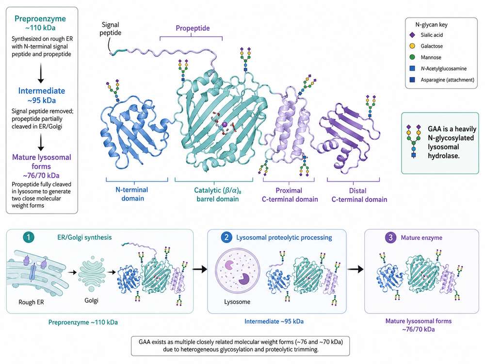
Figure 1. BioRender-style overview of human GAA architecture, glycosylation, and maturation from preproenzyme to mature lysosomal forms.
1. Overview of GAA Protein Architecture
Human GAA is a lysosomal glycoside hydrolase in the GH31 family. It is synthesized as a single polypeptide that contains an N-terminal signal peptide, a propeptide region, a catalytic core, and C-terminal structural elements that support substrate access and enzyme stability. The central catalytic region adopts a (β/α)8 barrel-like fold typical of several retaining glycosidases, while adjacent domains help form the substrate-binding surface and maintain the geometry of the active site.
From a functional perspective, GAA architecture can be viewed as a coordinated system: one part ensures biosynthetic trafficking, another part supports lysosomal processing, and another part performs catalysis on glycogen-derived substrates. This is especially relevant when choosing a GAA enzyme for biochemical, cellular, or translational studies, because seemingly small structural differences can alter assay performance.
| Structural Feature | General Role | Why It Matters Experimentally |
|---|---|---|
| Signal peptide and propeptide | Direct biosynthetic entry into the secretory pathway and enable early maturation steps | Influence correct secretion, precursor processing, and comparability between native and recombinant formats |
| Catalytic core | Contains the active site and key substrate-binding subsites | Determines specific activity, substrate preference, and pH responsiveness |
| N-linked glycans | Support folding, solubility, receptor recognition, and intracellular trafficking | Affect M6P content, uptake, and lysosomal delivery |
| C-terminal domains | Contribute to structural stability and substrate positioning | Relevant to stability studies and structure-function interpretation of variants |
2. Precursor and Mature Forms of GAA
GAA is initially translated as a preproenzyme of approximately 110 kDa. The signal peptide is removed co-translationally during entry into the endoplasmic reticulum, and the resulting precursor is glycosylated and trafficked through the Golgi apparatus. Proteolytic trimming during biosynthetic transit and, more importantly, after lysosomal delivery generates lower-molecular-weight mature forms that are typically reported around 95 kDa and then around 76/70 kDa.
These mature lysosomal forms are not simply smaller versions of the same molecule. Their processing is tied to conformational maturation and can improve access to the catalytic pocket or optimize enzyme function in the lysosomal environment. For this reason, studies that compare precursor versus mature alglucosidase alfa-like materials should document the molecular form being tested.
Molecular weight alone does not fully define GAA quality. Researchers should interpret precursor and mature bands in the context of glycosylation, proteolytic processing, and intracellular trafficking rather than assuming all forms are functionally equivalent.
3. Glycosylation and Lysosomal Targeting
Human GAA is a heavily N-glycosylated lysosomal enzyme, and these glycans are essential to its biology. They support protein folding in the secretory pathway, improve solubility, and provide the biochemical scaffold on which M6P can be added. M6P is then recognized by mannose-6-phosphate receptors, allowing the enzyme to be sorted toward lysosomes instead of remaining in the extracellular space.
For recombinant materials, glycosylation quality can be as important as purity or nominal activity. A preparation with adequate catalytic capacity in vitro may still show weak lysosomal correction in cells if glycan maturation or M6P density is suboptimal. This is one reason CHO-derived and glycoengineered recombinant human GAA materials are often characterized beyond routine SDS-PAGE and enzyme activity readouts.
| Glycosylation Topic | Functional Implication | Typical Research Readout |
|---|---|---|
| N-glycan occupancy | Supports secretion, folding, and structural integrity | Mass spectrometry, peptide mapping, mobility shifts, glycan profiling |
| M6P-bearing glycans | Promote receptor-mediated uptake and lysosomal targeting | CI-MPR binding, cellular uptake assays, lysosomal colocalization |
| Glycan heterogeneity | Creates lot-to-lot variation in trafficking and stability | HPAEC, LC-MS, glycopeptide analysis, cell-based potency |
| Terminal processing | May influence circulation, receptor interactions, and manufacturability | Comparative glycan mapping and stability testing |
4. Catalytic Domain and Active Site Function
The catalytic machinery of GAA is centered on conserved aspartate residues, commonly described as Asp518 and Asp616 in the mature human enzyme sequence framework. These residues work together in a retaining glycosidase mechanism to hydrolyze glycosidic bonds. The surrounding active-site pocket contains additional residues that help position carbohydrate substrates, stabilize transition states, and control catalytic efficiency under acidic lysosomal conditions.
Structural studies show that activity cannot be understood by the catalytic residues alone. The geometry of neighboring loops, the contour of substrate-binding subsites, and the local electrostatic environment all influence how efficiently glycogen-derived chains are engaged and cleaved. This has practical implications when designing assays or interpreting lower-than-expected activity of variant or recombinant enzymes.
5. Substrate Recognition of Glycogen
GAA does not encounter glycogen as a simple linear polymer. Lysosomal glycogen contains a mixture of α-1,4-linked glucose chains and α-1,6 branch points, meaning substrate recognition requires an extended surface that can bind multiple glucose units while orienting individual linkages for cleavage. Short fluorogenic substrates such as 4-MUG are useful for routine activity measurements, but they do not fully capture the way native glycogen engages the enzyme surface.
Substrate-binding subsites surrounding the active site help explain why one assay format may exaggerate or underestimate performance relative to another. When researchers compare wild-type enzyme, disease-associated variants, or engineered acid alpha-glucosidase products, substrate choice should be matched to the biological question.
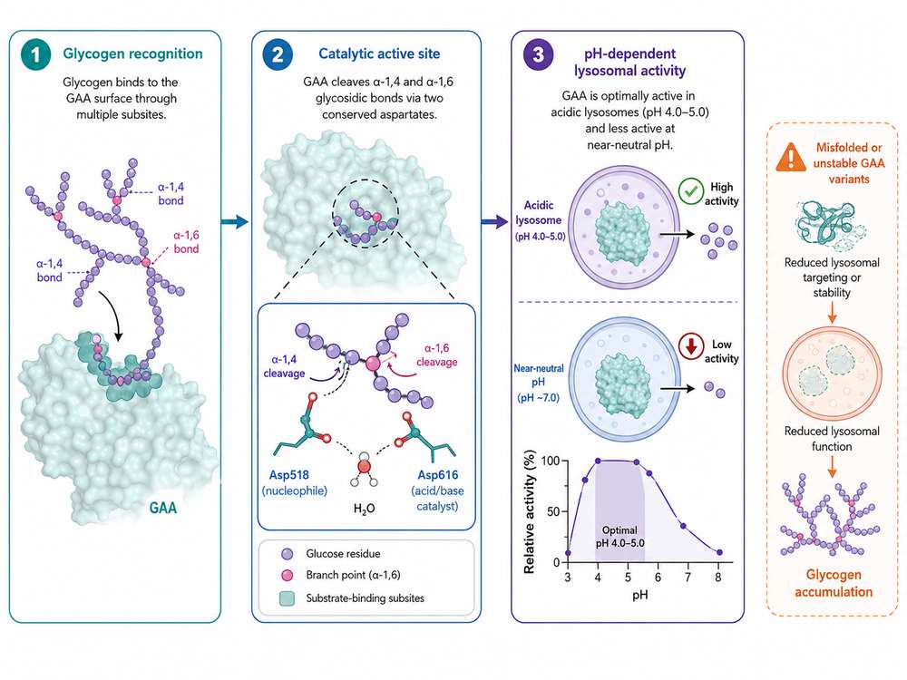
Figure 2. BioRender-style diagram linking glycogen recognition, catalytic active-site function, pH-dependent lysosomal activity, and the consequences of GAA misfolding or instability.
6. pH Dependence of Enzyme Activity
GAA is optimized for the acidic lysosomal lumen and typically shows highest activity in the approximate pH 4.0–5.0 range. This pH dependence reflects both catalytic chemistry and structural context. Protonation states within the active site affect how catalytic residues behave, while the acidic environment also supports substrate processing in a manner that differs from neutral extracellular or cytosolic conditions.
As a result, in vitro activity values are highly sensitive to buffer design. Assays run too far from the lysosomal pH window may underestimate the apparent function of otherwise active enzyme. Conversely, enzyme preparations that perform well only within a narrow range may be more vulnerable to environmental instability during handling, uptake, or intracellular trafficking.
7. Structural Instability and Misfolded GAA Variants
Many disease-associated GAA variants do not completely abolish catalysis; instead, they impair folding, reduce stability, alter trafficking, or increase susceptibility to degradation. This is one reason genotype–phenotype correlations in Pompe disease can be complex. A variant may retain partial catalytic competence in a purified assay but still fail in cells because too little properly folded enzyme reaches the lysosome.
From a research standpoint, misfolding can be detected indirectly through reduced secretion, increased aggregation, altered thermal stability, aberrant glycosylation patterns, or lower lysosomal processing efficiency. Structural biology therefore complements standard enzyme assays by explaining whether a loss-of-function phenotype is catalytic, conformational, or trafficking-related.
| Structural Problem | Likely Functional Outcome | Useful Experimental Approaches |
|---|---|---|
| Misfolding in ER | Poor secretion, intracellular retention, reduced mature enzyme formation | Western blot, pulse-chase, immunofluorescence, ER stress markers |
| Reduced M6P presentation | Weaker receptor-mediated uptake and lysosomal delivery | Uptake assays, receptor-blocking studies, glycan analysis |
| Active-site distortion | Low catalytic efficiency despite adequate expression | Kinetic analysis, substrate profiling, structural modeling |
| Aggregation or instability | Lower potency and variable assay reproducibility | SEC, DLS, thermal shift analysis, stress studies |
8. Implications for Recombinant Enzyme Design
Modern recombinant GAA design aims to optimize more than expression yield. The most informative quality framework considers correct folding, glycan occupancy, M6P content, proteolytic maturation, aggregation state, and specific activity together. In other words, a useful research-grade enzyme should not only be present and pure, but also structurally competent for lysosomal biology.
These considerations have driven glycoengineering approaches intended to improve receptor engagement and tissue uptake. Structural knowledge also guides formulation development, analytical characterization, and assay selection, especially when comparing biosimilar-like, investigational, or mechanistically modified GAA molecules.
9. Structure-Guided Research Applications
Structure-guided understanding of GAA supports a wide range of research activities, including variant interpretation, enzyme engineering, cell-based correction studies, and analytical comparability programs. For instance, researchers may combine enzyme kinetics with glycan profiling and uptake assays to distinguish a catalytically competent but trafficking-limited preparation from a globally unstable one.
In translational settings, structural information is also valuable when linking biochemical data to tissue outcomes. Better receptor targeting, more consistent lysosomal processing, or improved stability may translate into stronger glycogen clearance in skeletal muscle models. Therefore, selecting a recombinant human GAA reagent with well-documented structural attributes can substantially improve study interpretability.
| Research Goal | Most Relevant Structural Readouts | Typical Downstream Endpoint |
|---|---|---|
| Variant functional analysis | Folding, glycosylation, trafficking, catalytic activity | Pathogenicity assessment and mechanism assignment |
| Recombinant enzyme comparison | Purity, glycan profile, M6P, aggregation, specific activity | Material selection for cell or animal studies |
| Cellular correction studies | Receptor uptake, lysosomal delivery, mature form generation | Glycogen reduction and restoration of lysosomal function |
| Formulation and stability work | Conformational stability, stress sensitivity, lot consistency | Shelf-life and handling robustness |
10. Summary
GAA function emerges from a tightly connected structural system that includes domain organization, proteolytic maturation, glycosylation, M6P tagging, active-site architecture, and pH-adapted lysosomal catalysis. When any of these layers is compromised, enzyme performance can decline through reduced catalytic efficiency, unstable folding, weak intracellular targeting, or poor maturation.
For researchers developing assays, comparing enzyme preparations, or studying disease-associated variants, structure is therefore a practical guide rather than a purely descriptive topic. A well-characterized GAA enzyme should be evaluated through both biochemical and structural quality attributes to ensure that experimental conclusions reflect true lysosomal function.
In GAA research, function depends on structure at every stage—from folding and glycosylation to catalytic chemistry and lysosomal targeting. Structural characterization is therefore essential for reliable use of acid alpha-glucosidase in biochemical, cellular, and translational studies.
References
1. Roig-Zamboni, V., et al. (2017). Structure of human lysosomal acid α-glucosidase—a guide for the treatment of Pompe disease. Nature Communications, 8: 1111.
2. Moreland, R. J., et al. (2005). Lysosomal acid alpha-glucosidase consists of four different peptides processed from a single chain precursor. Journal of Biological Chemistry, 280(8): 6780-6791.
3. Wisselaar, H. A., et al. (1993). Structural and functional changes of lysosomal acid alpha-glucosidase during intracellular transport and maturation. Journal of Biological Chemistry, 268(30): 2223-2231.
4. Zhu, Y., et al. (2009). Glycoengineered acid alpha-glucosidase with improved efficacy at correcting the metabolic aberrations and motor function deficits in a mouse model of Pompe disease. Molecular Therapy, 17(6): 954-963.
5. Maga, J. A., et al. (2013). Glycosylation-independent lysosomal targeting of acid alpha-glucosidase enhances muscle glycogen clearance in Pompe mice. Journal of Biological Chemistry, 288(3): 1428-1438.
6. Kishnani, P. S., et al. (2007). A retrospective, multinational, multicenter study on the natural history of infantile-onset Pompe disease. Journal of Pediatrics, 148(5): 671-676.
7. Kishnani, P. S., et al. (2006). Pompe disease diagnosis and management guideline. Genetics in Medicine, 8(5): 267-288.
8. van der Ploeg, A. T., & Reuser, A. J. J. (2008). Pompe's disease. The Lancet, 372(9646): 1342-1353.
9. Winchester, B., et al. (2008). Methods for a prompt and reliable laboratory diagnosis of Pompe disease: report from an international consensus meeting. Molecular Genetics and Metabolism, 93(3): 275-281.
10. Lim, J. A., et al. (2014). Defects in calcium homeostasis and mitochondria can be reversed by restoring autophagy in Pompe disease. Autophagy, 11(2): 385-402.
11. Do, H. T. T., et al. (2019). Crystal structure of human lysosomal acid alpha-glucosidase with acarbose and implications for catalysis. Protein Science, 28(9): 1630-1642.
12. Schoser, B., et al. (2024). Pompe disease: unmet needs and emerging therapies. Molecular Genetics and Metabolism, 142(4): 108513.
13. Parenti, G., et al. (2014). Pharmacological chaperone therapy for lysosomal storage diseases. Future Medicinal Chemistry, 6(3): 307-318.
14. EMA. Myozyme (alglucosidase alfa) product information. European Medicines Agency.