
Pompe Disease Research: Linking GAA Deficiency, Lysosomal Glycogen Accumulation, and Enzyme Therapy
From GAA Gene Variants and Muscle Pathology to Recombinant Enzyme Evaluation and Translational Models
Abstract
Pompe disease, also called glycogen storage disease type II, is an autosomal recessive lysosomal storage disorder caused by deficient acid alpha-glucosidase activity. In healthy cells, this enzyme hydrolyzes glycogen within the acidic lysosomal compartment. Pathogenic variants in the GAA gene can reduce enzyme synthesis, folding, trafficking, catalytic activity, or lysosomal stability, allowing glycogen to accumulate progressively in skeletal muscle, cardiac muscle, and respiratory muscle.
The resulting pathology is not explained by storage alone. Enlarged glycogen-filled lysosomes, impaired autophagic flux, disruption of the contractile apparatus, altered vesicular trafficking, mitochondrial stress, inflammation, and loss of muscle fiber integrity may all contribute to disease progression. Recombinant human GAA has therefore become both a therapeutic modality and a central experimental reagent for studying receptor-mediated uptake, lysosomal delivery, glycogen clearance, immune responses, and tissue-specific treatment barriers. This article connects molecular defects with cellular pathology, diagnostic research markers, translational models, and enzyme therapy development.
Pompe disease research, GAA deficiency, glycogen storage disease type II, acid alpha-glucosidase deficiency, alglucosidase alfa, lysosomal storage disorder
1. Overview of Pompe Disease
Pompe disease occupies a distinctive position among glycogen storage disorders because the primary defect lies in lysosomal, rather than cytosolic, glycogen degradation. The disease results from reduced or absent activity of acid alpha-glucosidase, also known as GAA or acid maltase. Glycogen continues to enter the lysosomal system through autophagic and vesicular pathways, but inadequate hydrolysis prevents efficient recycling to free glucose.
Disease severity spans a continuum. Classic infantile-onset Pompe disease is typically associated with profound enzyme deficiency, rapidly progressive hypertrophic cardiomyopathy, generalized hypotonia, and respiratory compromise. Late-onset Pompe disease usually retains more residual GAA activity and presents with progressive axial, limb-girdle, diaphragmatic, and respiratory muscle weakness. This phenotypic range makes Pompe disease a useful model for examining how residual lysosomal enzyme activity, variant type, tissue biology, and treatment timing interact.
How does a quantitative reduction in GAA activity become a multisystem muscle disorder, and which steps in enzyme delivery most strongly determine therapeutic correction?
2. Genetic Basis: Mutations in the GAA Gene
The human GAA gene is located on chromosome 17q25.3 and encodes a 952-amino-acid precursor that enters the secretory pathway before mannose-6-phosphate-dependent delivery to lysosomes. Hundreds of disease-associated variants have been described, including missense, nonsense, splice-site, frameshift, insertion/deletion, and larger rearrangement events. These variants can affect transcription, mRNA splicing, protein folding, catalytic residues, glycosylation, intracellular transport, proteolytic maturation, or stability in the lysosome.
Genotype-phenotype relationships are informative but not absolute. Variants that abolish protein production or catalytic function are commonly associated with severe early disease, whereas hypomorphic alleles may permit enough residual activity to delay onset. However, modifier genes, tissue-specific expression, immune status, and treatment history can alter the observed phenotype. For experimental studies, variant characterization should therefore be paired with direct measurements of GAA activity and intracellular processing.
| Variant Effect | Possible Molecular Consequence | Recommended Research Readout |
|---|---|---|
| Loss of transcript or translation | Minimal or absent GAA precursor | qPCR, transcript analysis, immunoblotting, enzyme activity |
| Protein misfolding | ER retention, degradation, reduced secretion | Cellular localization, secretion assay, proteostasis markers |
| Trafficking defect | Reduced mannose-6-phosphate receptor-mediated lysosomal delivery | Uptake assay, lysosomal colocalization, maturation profile |
| Catalytic-site alteration | Protein present but substrate hydrolysis impaired | Specific activity, kinetic analysis, glycogen degradation assay |
| Partial-function allele | Residual activity with delayed substrate accumulation | Low-range activity assay, longitudinal glycogen and phenotype analysis |
3. Lysosomal Glycogen Accumulation and Cellular Dysfunction
In GAA-deficient cells, glycogen accumulates within lysosomes because its α-1,4 and α-1,6 glycosidic linkages cannot be degraded efficiently in the acidic lumen. Lysosomes enlarge, redistribute, and may occupy substantial regions of the muscle fiber. As storage progresses, membrane integrity and organelle communication can deteriorate, while glycogen and vesicular material interfere with the ordered architecture required for contraction.
Autophagic dysfunction is a major secondary feature. Autophagosomes and late endosomal structures can accumulate, particularly in skeletal muscle, creating autophagic buildup that impedes trafficking of both endogenous GAA and therapeutic enzyme. Additional reported mechanisms include altered calcium handling, mitochondrial dysfunction, oxidative stress, inflammatory signaling, defective proteostasis, and neuromuscular involvement. These findings support a multi-endpoint model in which substrate storage initiates a broader network of cellular injury.
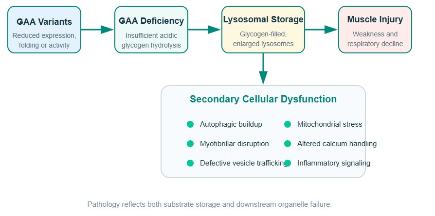
Figure 1: Mechanistic progression from GAA deficiency to lysosomal storage, secondary cellular dysfunction, and tissue pathology.
4. Infantile-Onset and Late-Onset Pompe Disease
Infantile-onset and late-onset forms share the same biochemical defect but differ in residual enzyme activity, rate of glycogen accumulation, dominant tissue involvement, and clinical tempo. These categories are useful for study design, although intermediate and atypical phenotypes occur.
| Feature | Classic Infantile-Onset Pompe Disease | Late-Onset Pompe Disease |
|---|---|---|
| Typical onset | First months of life | Childhood through adulthood |
| Residual GAA activity | Usually very low or absent | Usually measurable but below normal |
| Cardiac involvement | Prominent hypertrophic cardiomyopathy | Usually limited or absent |
| Skeletal muscle phenotype | Generalized hypotonia and weakness | Progressive axial and limb-girdle weakness |
| Respiratory involvement | Early and rapidly progressive | May be disproportionate to limb weakness |
| Research emphasis | Early intervention, immune tolerance, cardiac correction | Skeletal-muscle delivery, long-term function, respiratory endpoints |
Cross-reactive immunologic material status is particularly important in infantile studies. Individuals who produce no endogenous GAA protein may be at greater risk of developing high sustained anti-drug antibody responses to recombinant enzyme. Experimental programs evaluating recombinant human GAA should therefore consider both biochemical severity and immunological context.
5. Muscle and Respiratory Pathology
5.1 Skeletal Muscle
Skeletal muscle is a major site of glycogen storage and a difficult target for enzyme delivery. Glycogen-filled lysosomes, autophagic buildup, fiber-type differences, and relatively limited receptor-mediated uptake can reduce correction. Histological findings may include vacuolization, disrupted myofibrils, variable fiber size, and progressive replacement by noncontractile tissue.
5.2 Cardiac Muscle
Cardiac involvement is especially prominent in classic infantile disease. Cardiomyocyte lysosomal expansion and glycogen storage can produce marked ventricular hypertrophy and impaired cardiac performance. The strong cardiac response often observed after early enzyme therapy illustrates that tissue access and treatment timing are major determinants of reversibility.
5.3 Respiratory System
Weakness of the diaphragm, intercostal muscles, and accessory respiratory muscles can cause sleep-disordered breathing, hypoventilation, ineffective cough, and progressive respiratory insufficiency. Respiratory decline may precede severe ambulatory impairment in late-onset disease, making diaphragm-focused models and pulmonary function endpoints essential in translational research.
6. Diagnostic Research Markers: GAA Activity and Glycogen Storage
Low GAA activity is the defining biochemical marker, but robust diagnosis and disease modeling require orthogonal confirmation. Dried blood spots are useful for screening, whereas leukocytes, fibroblasts, muscle tissue, or other validated cell sources can support confirmatory analysis. Genetic testing identifies pathogenic GAA variants and helps distinguish true deficiency from assay artifacts or pseudodeficiency alleles.
| Marker or Method | What It Measures | Strength | Research Limitation |
|---|---|---|---|
| GAA activity assay | Hydrolysis of fluorogenic or natural substrate at acidic pH | Direct biochemical evidence of deficiency | Artificial substrates may not predict lysosomal glycogen clearance |
| GAA genotyping | Pathogenic or likely pathogenic variants | Confirms molecular basis and supports family studies | Variant interpretation may be uncertain for novel alleles |
| Glycogen quantification | Total or tissue-specific substrate burden | Measures disease substrate directly | Total content does not reveal subcellular localization |
| Urinary Glc4 | Glucose tetrasaccharide associated with glycogen breakdown | Noninvasive longitudinal biomarker | Not fully specific and influenced by age or disease context |
| CK and muscle enzymes | Muscle injury or membrane leakage | Accessible supportive marker | May be normal or only mildly elevated |
| Imaging and functional tests | Muscle composition, strength, mobility, pulmonary function | Links biology with tissue performance | Changes may lag behind biochemical correction |
For mechanistic studies, the strongest design combines enzyme activity, genetic confirmation, glycogen burden, lysosomal imaging, autophagy markers, and tissue function. This approach avoids overinterpreting a single biochemical endpoint.
7. Role of Recombinant GAA in Therapeutic Development
Enzyme replacement therapy uses recombinant GAA to restore lysosomal glycogen hydrolysis. After intravenous administration or addition to cell culture, the enzyme must remain stable, bind cell-surface cation-independent mannose-6-phosphate receptors, undergo endocytosis, reach acidic endosomes and lysosomes, mature proteolytically, and access stored glycogen. Failure at any step can limit efficacy even when catalytic activity is high in a cell-free assay.
Alglucosidase alfa established the feasibility of recombinant enzyme replacement and remains an important benchmark for comparative studies. Current research investigates higher M6P content, glycoengineering, receptor-targeting motifs, pharmacological chaperones, altered dosing, immune modulation, and gene-based delivery. These strategies aim to improve skeletal-muscle exposure, reduce antibody-mediated loss of activity, and achieve more durable lysosomal correction.
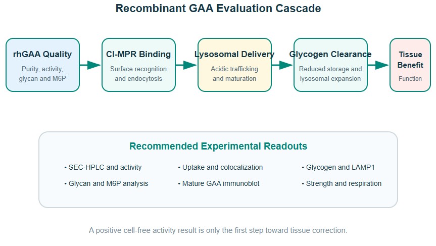
Figure 2: Experimental pathway linking recombinant GAA quality and uptake to lysosomal correction and muscle-function outcomes.
Recombinant enzyme potency should be evaluated as a delivery-and-function cascade: glycan quality → receptor binding → internalization → lysosomal maturation → glycogen clearance → tissue recovery.
8. Preclinical and Translational Research Models
No single model reproduces the full clinical spectrum. A tiered strategy is preferable, beginning with biochemical and cellular assays, progressing to patient-derived systems, and then testing whole-body distribution and muscle function in animal models.
| Model | Primary Use | Representative Endpoints | Key Limitation |
|---|---|---|---|
| Purified enzyme assays | Catalytic potency and stability | Specific activity, pH profile, kinetics, thermal stability | No information on cellular uptake or trafficking |
| GAA-deficient fibroblasts | Uptake and lysosomal delivery | Internalization, maturation, glycogen reduction | Limited representation of contractile muscle biology |
| Myoblasts and differentiated myotubes | Skeletal-muscle correction | Lysosomal glycogen, autophagy, morphology, contraction | Culture conditions can alter receptor expression |
| Patient-derived iPSC muscle or 3D tissue | Human genotype-specific disease modeling | Fiber maturation, force, organelle pathology, treatment response | Complex production and batch variability |
| Gaa knockout mouse | Systemic biodistribution and functional efficacy | Tissue glycogen, strength, respiratory function, histology | Immune and disease-course differences from humans |
| Large-animal or advanced translational model | Dose scaling and delivery validation | Pharmacokinetics, organ distribution, safety, device compatibility | Cost, availability, and ethical constraints |
Model selection should match the development question. Glycoengineering studies require receptor-binding and uptake assays; immune-response studies require antibody and tolerance endpoints; respiratory programs need diaphragm and ventilation measures; and gene therapy programs must evaluate expression durability, biodistribution, and vector-associated immunity.
9. Challenges in Studying Pompe Disease Mechanisms
- Residual activity is difficult to interpret: very low activity values can be assay-dependent, and small differences may have large phenotypic consequences.
- Artificial substrate assays can overestimate correction: efficient cleavage of a small fluorogenic substrate does not guarantee uptake or degradation of lysosomal glycogen.
- Muscle is heterogeneous: fiber type, receptor abundance, perfusion, autophagic burden, and disease stage influence treatment response.
- Autophagic buildup can obstruct delivery: therapeutic enzyme may enter cells but fail to distribute efficiently through diseased fibers.
- Immune responses confound exposure: anti-GAA antibodies may alter pharmacokinetics, uptake, safety, and long-term efficacy.
- Biochemical and functional recovery are asynchronous: glycogen reduction may occur before measurable improvement in strength or respiratory performance.
- Model systems incompletely reproduce human disease: species-specific receptor biology and immune responses complicate translation.
These challenges favor experimental designs that combine matched controls, multiple time points, orthogonal readouts, and careful characterization of the GAA enzyme preparation, including purity, aggregation, glycosylation, M6P content, endotoxin, and stability.
10. Research Outlook
Pompe disease research is shifting from simple enzyme supplementation toward integrated correction of delivery, immunity, autophagy, and tissue function. Next-generation recombinant enzymes are being designed to improve receptor engagement and skeletal-muscle uptake. Gene therapy and liver-directed expression strategies seek sustained systemic enzyme supply, while genome editing and mRNA approaches may offer additional routes to durable correction. Combination strategies may be required when advanced muscle pathology limits the effect of substrate reduction alone.
New biomarkers and quantitative imaging methods are also expanding the ability to monitor tissue glycogen and treatment response. High-content microscopy, spatial omics, patient-derived 3D muscle, organ-on-chip systems, and noninvasive glycogen-sensitive imaging may help identify which cellular abnormalities are reversible and when intervention is most effective.
The most informative future studies will link GAA genotype and biochemical activity to lysosomal trafficking, glycogen clearance, autophagic recovery, muscle mechanics, respiratory performance, and immune response. Within this framework, recombinant acid alpha-glucosidase remains both a foundational therapy and a powerful research tool for dissecting the barriers between enzyme exposure and meaningful tissue correction.
Success will depend not only on delivering more GAA, but on delivering the right molecular form to the right muscle compartments early enough to restore lysosomal and contractile function.
References
1. van der Ploeg, A. T., & Reuser, A. J. J. (2008). Pompe's disease. The Lancet, 372(9646): 1342-1353.
2. Kishnani, P. S., et al. (2006). Pompe disease diagnosis and management guideline. Genetics in Medicine, 8(5): 267-288.
3. Leslie, N. D., & Tinkle, B. T. Pompe Disease. GeneReviews®. University of Washington, Seattle.
4. Moreland, R. J., et al. (2005). Lysosomal acid alpha-glucosidase consists of four different peptides processed from a single-chain precursor. Journal of Biological Chemistry, 280(8): 6780-6791.
5. Roig-Zamboni, V., et al. (2017). Structure of human lysosomal acid α-glucosidase—a guide for the treatment of Pompe disease. Nature Communications, 8: 1111.
6. Fukuda, T., et al. (2006). Dysfunction of endocytic and autophagic pathways in a lysosomal storage disease. Annals of Neurology, 59(4): 700-708.
7. Raben, N., et al. (2010). Autophagy in skeletal muscle: implications for Pompe disease. Autophagy, 6(7): 847-856.
8. Sánchez-Porras, V., & Echeverri-Peña, O. Y. (2023). From acid alpha-glucosidase deficiency to autophagy: understanding the bases of Pompe disease. International Journal of Molecular Sciences, 24(15): 12481.
9. Winchester, B., et al. (2008). The molecular basis of glycogen storage disease type II (Pompe disease). Human Mutation, 29(6): 733-747.
10. Toscano, A., & Schoser, B. (2013). Enzyme replacement therapy in late-onset Pompe disease: a systematic literature review. Journal of Neurology, 260: 951-959.
11. van den Hout, J. M. P., et al. (2000). Recombinant human alpha-glucosidase from rabbit milk in Pompe patients. The Lancet, 356(9227): 397-398.
12. Zhu, Y., et al. (2009). Glycoengineered acid alpha-glucosidase with improved efficacy in a mouse model of Pompe disease. Molecular Therapy, 17(6): 954-963.
13. Niñerola-Baizán, A., et al. (2020). Enzymatic diagnosis of Pompe disease: lessons from 28 years of experience. European Journal of Human Genetics, 29: 434-446.
14. Wang, J., et al. (2021). Three-dimensional tissue-engineered human skeletal muscle model of Pompe disease. Communications Biology, 4: 524.
15. Schoser, B., et al. (2024). Pompe disease: unmet needs and emerging therapies. Molecular Genetics and Metabolism, 142(4): 108513.
16. Lim, J. A., et al. (2024). Failure of autophagy in Pompe disease. Biomolecules, 14(5): 573.