
Acid Alpha-Glucosidase FAQ: GAA Function, Assays, Applications, and Research Use
Clear Answers for Recombinant GAA Selection, Experimental Design, and Data Interpretation
Overview
Acid alpha-glucosidase is central to lysosomal glycogen degradation and is widely studied in Pompe disease biology, recombinant enzyme characterization, enzyme replacement research, and cell-based lysosomal delivery models. This FAQ summarizes practical distinctions between native GAA, recombinant GAA, and alglucosidase alfa; outlines commonly used activity and uptake assays; and highlights factors that can affect experimental reproducibility.
acid alpha-glucosidase, recombinant GAA, alglucosidase alfa, Pompe disease research, lysosomal glycogen degradation, GAA activity assay, mannose-6-phosphate receptor-mediated uptake
| Term | Meaning | Typical Research Context |
|---|---|---|
| GAA | Acid alpha-glucosidase; the lysosomal enzyme encoded by the human GAA gene | Disease biology, enzyme function, genetics, assay development |
| rhGAA | Recombinant human acid alpha-glucosidase | Biochemical testing, uptake studies, comparability, engineering |
| Alglucosidase alfa | A recombinant human GAA therapeutic protein | ERT research, reference comparisons, translational studies |
| M6P | Mannose-6-phosphate glycan signal recognized by M6P receptors | Cellular uptake and lysosomal targeting |
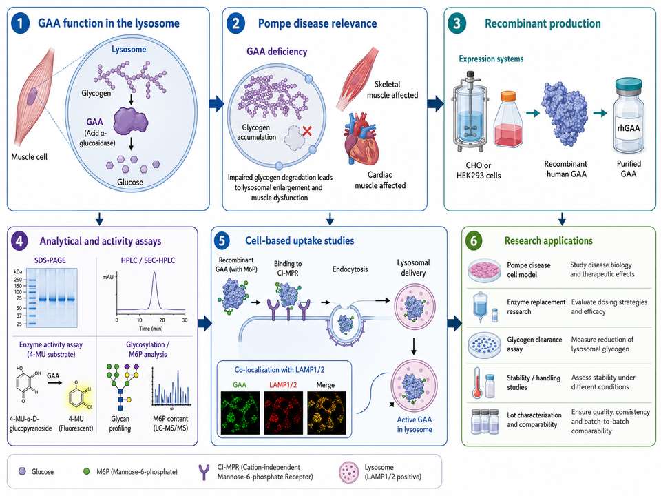
Figure 1: Overview of GAA biology and research use, connecting lysosomal glycogen degradation with recombinant production, analytical testing, cellular uptake, and experimental applications.
1. What Is Acid Alpha-Glucosidase?
Acid alpha-glucosidase, commonly abbreviated as GAA, is a lysosomal glycoside hydrolase that breaks down glycogen within the acidic environment of the lysosome. It hydrolyzes both alpha-1,4 and alpha-1,6 glycosidic linkages, ultimately generating free glucose. The enzyme is synthesized as a glycosylated precursor, trafficked through the secretory pathway, and processed into mature lysosomal forms.
In research, Acid alpha-glucosidase may refer to the endogenous human enzyme, a purified native preparation, or a recombinant protein. These materials are not automatically interchangeable because expression system, glycosylation, processing, purity, formulation, and activity can differ.
2. What Is the Difference Between GAA and Alglucosidase Alfa?
GAA is the general name for the human enzyme and the protein product of the GAA gene. Alglucosidase alfa is a recombinant human GAA therapeutic protein developed as an exogenous source of lysosomal GAA. Therefore, all alglucosidase alfa is recombinant GAA, but not every recombinant GAA preparation should be described as alglucosidase alfa.
| Feature | GAA | Alglucosidase Alfa |
|---|---|---|
| Definition | Human lysosomal acid alpha-glucosidase enzyme | Defined recombinant therapeutic form of human GAA |
| Source | Endogenous, native purified, or recombinant | Manufactured recombinant protein |
| Research Use | Mechanistic, biochemical, genetic, or cell biology studies | ERT-related studies and benchmark comparisons |
| Interchangeability | Depends on product attributes and intended application | Should be identified by its specific product characteristics |
When documenting an experiment, report the exact material used, including supplier, catalog number, lot number, expression system, specific activity, formulation, and storage history.
3. What Role Does GAA Play in Glycogen Degradation?
Cytosolic glycogenolysis and lysosomal glycogen degradation are distinct but complementary processes. GAA functions inside lysosomes, where it cleaves glycogen delivered through lysosomal and autophagic pathways. Functional GAA activity prevents excessive expansion of glycogen-filled lysosomes and supports normal lysosomal homeostasis.
| Stage | Role of GAA | Possible Experimental Readout |
|---|---|---|
| Lysosomal delivery | GAA reaches the lysosomal compartment | Co-localization with LAMP1/LAMP2 or LysoTracker |
| Substrate hydrolysis | Cleaves alpha-linked glucose residues in glycogen | 4-MU substrate activity or glucose-release assay |
| Glycogen reduction | Decreases lysosomal glycogen burden | PAS staining, glycogen assay, microscopy |
| Cellular correction | Supports improved lysosomal structure and function | Organelle morphology, autophagy markers, functional endpoints |
4. Why Is GAA Important in Pompe Disease Research?
Pompe disease is caused by deficient or absent GAA activity, leading to lysosomal glycogen accumulation, particularly in skeletal, respiratory, and cardiac muscle. GAA research therefore spans disease mechanism, genotype–phenotype relationships, enzyme replacement, receptor-mediated uptake, immune response, biomarker development, gene therapy, and combination strategies.
A recombinant GAA study may focus on a single step—such as catalytic activity—or on an integrated sequence that includes receptor binding, uptake, lysosomal localization, enzyme recovery, glycogen clearance, and cellular phenotype correction.
5. How Is Recombinant Human GAA Commonly Produced?
Recombinant human GAA is commonly expressed in mammalian systems because the enzyme requires complex folding, disulfide-bond formation, N-linked glycosylation, and lysosomal-targeting glycans. CHO cells are widely used, while HEK293 cells and other engineered systems may be selected for research production or glycoengineering studies.
| Expression System | Potential Advantages | Points to Verify |
|---|---|---|
| CHO cells | Established scalable mammalian platform; complex glycosylation | M6P content, sialylation, processing, host-cell impurities |
| HEK293 cells | Flexible transient or stable expression; useful for research variants | Lot scale, glycan profile, precursor/mature form distribution |
| Alternative eukaryotic systems | Potentially distinct glycan engineering or production economics | Human compatibility of glycans and receptor-mediated uptake |
| Cell-free or non-mammalian systems | Rapid prototyping in selected applications | Folding, glycosylation, activity, and lysosomal-targeting competence |
For uptake-focused studies, the expression platform alone is not sufficient information. Researchers should review glycosylation, exposed M6P, receptor binding, cellular internalization, and lysosomal function data.
6. What Assays Are Used to Measure GAA Activity?
The most common biochemical method uses 4-methylumbelliferyl-alpha-D-glucopyranoside (4-MU-alpha-Glc). GAA hydrolysis releases fluorescent 4-methylumbelliferone, which is measured after stopping or alkalinizing the reaction. Natural or glycogen-like substrates, glucose-release methods, LC-based assays, and cell-based functional readouts can provide complementary information.
| Assay Type | Typical Readout | Strength | Key Limitation |
|---|---|---|---|
| 4-MU fluorometric assay | Fluorescence proportional to substrate hydrolysis | Sensitive, rapid, plate-compatible | Artificial substrate may not reflect all functional properties |
| Colorimetric substrate assay | Absorbance change | Accessible instrumentation | Often lower sensitivity |
| Natural substrate assay | Glucose or glycogen breakdown | Closer to physiological function | More complex sample preparation and interference control |
| Cellular uptake/activity assay | Intracellular GAA activity after treatment | Integrates uptake and catalytic function | Influenced by cell model and receptor expression |
| Glycogen-clearance assay | Reduction of cellular glycogen | Direct functional outcome | Longer assay and greater biological variability |
Use a biochemical activity assay to confirm catalytic competence, then add uptake, lysosomal localization, and glycogen-clearance assays when the research question concerns cellular delivery or functional correction.
7. Why Does Glycosylation Matter for GAA?
GAA is a heavily glycosylated lysosomal enzyme. Its N-glycan profile can influence folding, stability, serum clearance, tissue distribution, receptor binding, internalization, and lysosomal delivery. Mannose-6-phosphate-bearing glycans are particularly important because they support binding to cation-independent mannose-6-phosphate receptor pathways involved in cellular uptake.
| Glycan Attribute | Potential Effect | Useful Method |
|---|---|---|
| M6P content and accessibility | CI-MPR binding and cellular uptake | HPAEC-PAD, LC-MS, receptor-binding assay |
| Sialylation | Circulatory persistence and reduced off-target hepatic clearance | Released-glycan LC-MS, sialic acid analysis |
| High-mannose structures | May alter receptor interactions and biodistribution | Glycopeptide mapping |
| Site occupancy | Can affect folding, stability, and comparability | Peptide/glycopeptide LC-MS |
8. How Should Recombinant GAA Be Stored and Handled?
Storage and handling requirements depend on the exact formulation. Researchers should follow the product-specific datasheet rather than applying a single universal condition. In general, recombinant enzymes are sensitive to repeated freeze–thaw cycles, adsorption to surfaces at low concentration, vigorous mixing, prolonged room-temperature exposure, incompatible pH, and microbial contamination.
| Handling Step | Recommended Practice | Risk if Poorly Controlled |
|---|---|---|
| Receipt | Verify cold chain, vial integrity, label, and appearance | Unrecognized temperature excursion or damage |
| Reconstitution | Use compatible buffer; swirl or invert gently | Foaming, aggregation, incomplete dissolution |
| Aliquoting | Prepare small single-use aliquots in low-binding tubes | Repeated freeze–thaw and adsorption loss |
| Dilution | Use validated buffer and carrier protein where appropriate | Activity loss at low concentration |
| Documentation | Record date, temperature, freeze–thaw count, and operator | Untraceable assay drift |
Additional practical guidance is provided in the recombinant GAA product information and corresponding lot documentation.
9. Can GAA Be Used in Cell-Based Uptake Studies?
Yes. Recombinant GAA is frequently evaluated in Pompe patient fibroblasts, myoblasts, differentiated myotubes, GAA-deficient engineered cells, and other tissue-relevant models. Uptake is commonly assessed through intracellular enzyme activity, labeled-protein internalization, receptor competition, immunofluorescence, lysosomal co-localization, western blotting, or glycogen clearance.
| Experimental Question | Recommended Readout | Useful Control |
|---|---|---|
| Is uptake receptor-dependent? | Intracellular GAA after treatment | Excess M6P or receptor-blocking condition |
| Does GAA reach lysosomes? | Co-localization with LAMP1/LAMP2 | Untreated cells and secondary-only imaging control |
| Is internalized GAA active? | Cell-lysate 4-MU activity assay | Heat-inactivated enzyme or no-enzyme control |
| Does treatment reduce substrate? | PAS staining or biochemical glycogen assay | Wild-type, untreated deficient, and positive treatment controls |
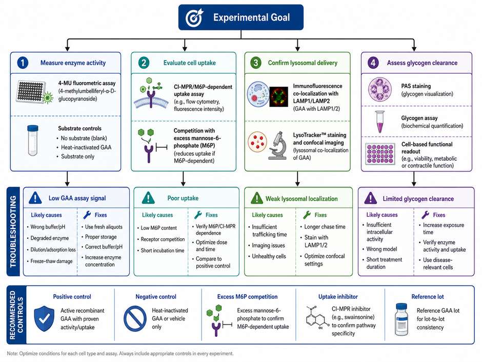
Figure 2: Decision map for selecting recombinant GAA assays and troubleshooting low activity, poor uptake, weak lysosomal localization, or limited glycogen clearance.
10. What Are Common Causes of Low GAA Assay Signal?
Low signal does not always indicate an inactive enzyme. The root cause may involve protein loss, assay chemistry, instrument settings, substrate quality, incubation conditions, cell uptake, receptor availability, or normalization errors.
| Observation | Possible Cause | Troubleshooting Step |
|---|---|---|
| Low signal in purified-enzyme assay | Wrong pH, degraded substrate, inactive enzyme, dilution error | Run fresh standards, verify buffer and substrate, compare a reference lot |
| High variability between wells | Pipetting error, uneven timing, bubbles, plate-edge effects | Use calibrated pipettes, synchronized addition, and consistent plate layout |
| Good in vitro activity but low cellular uptake | Low M6P exposure, low receptor expression, short uptake period | Measure receptor level, extend dose/time matrix, add M6P competition control |
| Uptake observed but limited glycogen reduction | Insufficient lysosomal delivery, inadequate dose, short treatment window | Confirm co-localization, intracellular activity, and longer functional follow-up |
| Loss after dilution | Surface adsorption or unstable low-concentration formulation | Use low-binding plasticware and validated carrier/stabilizer |
| Signal higher than expected | Endogenous enzyme, off-target glucosidase, substrate background | Use selective conditions, blank correction, and deficient-cell controls |
11. What Should Researchers Consider When Selecting Recombinant GAA?
Selection should be based on the intended application rather than on concentration or nominal purity alone. A preparation suitable for a basic biochemical assay may not be suitable for receptor-mediated uptake, lysosomal delivery, or translational comparison.
| Selection Criterion | Why It Matters | Evidence to Request |
|---|---|---|
| Species and sequence | Determines relevance and immunoreactivity | Sequence, accession, construct boundaries |
| Expression system | Influences folding and glycosylation | Host cell and production description |
| Purity and aggregation | Affects activity, background, and reproducibility | SDS-PAGE, SEC-HPLC, monomer percentage |
| Specific activity | Supports dose normalization and lot comparison | Assay method, units, substrate, pH, temperature |
| Glycosylation/M6P | Critical for uptake-focused studies | Glycan profile, M6P analysis, receptor or cell uptake data |
| Endotoxin | Important for immune and cell-based studies | Lot-specific endotoxin result |
| Formulation | Can affect stability and assay compatibility | Buffer composition and excipient information |
| Lot documentation | Supports reproducibility | Certificate of analysis and handling instructions |
For catalytic studies, prioritize specific activity and assay compatibility. For cell uptake studies, add M6P-related attributes and cellular internalization data. For immune or translational studies, also consider endotoxin, aggregation, formulation, and lot comparability.
12. Where Can Users Find Related GAA Resources?
The integrated alglucosidase alfa and recombinant Acid alpha-glucosidase page provides a central entry point for product information and related GAA research content. Useful supporting topics include:
- Cell-based evaluation of recombinant GAA uptake and lysosomal function
- Mannose-6-phosphate receptor-mediated delivery of Acid alpha-glucosidase
- Next-generation GAA therapeutic strategies
- Recombinant GAA immunogenicity considerations
- Stability and handling of recombinant Acid alpha-glucosidase
- Analytical characterization of recombinant GAA
When using any resource, distinguish educational research information from clinical instructions. Clinical dosing, administration, and patient management decisions must rely on current approved labeling and qualified healthcare guidance.
References
1. Hirschhorn, R., & Reuser, A. J. J. (2001). Glycogen storage disease type II: Acid alpha-glucosidase deficiency. In The Metabolic and Molecular Bases of Inherited Disease.
2. van der Ploeg, A. T., & Reuser, A. J. J. (2008). Pompe's disease. Lancet, 372(9646), 1342–1353.
3. Martiniuk, F., et al. (1990). Isolation of a cDNA for human acid alpha-glucosidase and detection of genetic heterogeneity in Pompe disease. PNAS, 87(7), 2478–2482.
4. Van Hove, J. L. K., et al. (1996). High-level production of recombinant human lysosomal acid alpha-glucosidase in Chinese hamster ovary cells. PNAS, 93(1), 65–70.
5. Zhu, Y., et al. (2009). Glycoengineered acid alpha-glucosidase with improved efficacy in a mouse model of Pompe disease. Molecular Therapy, 17(6), 954–963.
6. Cardone, M., et al. (2008). Abnormal mannose-6-phosphate receptor trafficking impairs recombinant alpha-glucosidase uptake in Pompe disease fibroblasts. Pathogenetics, 1, 6.
7. U.S. Food and Drug Administration. MYOZYME/LUMIZYME prescribing information: alglucosidase alfa.
8. ICH Q6B. Specifications: Test Procedures and Acceptance Criteria for Biotechnological/Biological Products.
9. Sánchez-Porras, V., & Echeverri-Peña, O. Y. (2023). From acid alpha-glucosidase deficiency to autophagy: Understanding the bases of Pompe disease. International Journal of Molecular Sciences, 24(15), 12481.