
Acid Alpha-Glucosidase Biology: Mechanism, Structure, and Role in Lysosomal Glycogen Degradation
From GAA Biosynthesis and Lysosomal Targeting to Glycogen Hydrolysis and Pompe Disease Research
Abstract
Acid alpha-glucosidase (GAA), also known as lysosomal alpha-glucosidase or acid maltase, is a glycoside hydrolase that converts lysosomal glycogen into free glucose. Unlike cytosolic glycogenolysis, which is mediated mainly by glycogen phosphorylase and the cytosolic debranching enzyme, lysosomal glycogen degradation depends on the delivery, maturation, and catalytic activity of GAA in an acidic compartment. This pathway represents a quantitatively smaller but biologically indispensable route for glycogen turnover, particularly in long-lived and metabolically demanding muscle cells.
Loss or reduction of GAA activity causes lysosomal glycogen accumulation and underlies glycogen storage disease type II, commonly known as Pompe disease. The resulting pathology extends beyond simple substrate storage: enlarged lysosomes, impaired autophagic flux, altered vesicular trafficking, myofibrillar disruption, and progressive dysfunction of skeletal, cardiac, and respiratory muscle may all contribute to the phenotype. Consequently, acid alpha-glucosidase is central to studies of lysosomal biology, muscle metabolism, disease modeling, enzyme replacement, and next-generation delivery strategies.
Acid alpha-glucosidase, GAA enzyme, lysosomal glycogen degradation, glycogen storage disease type II, Pompe disease research, recombinant GAA, alglucosidase alfa
1. Introduction to Acid Alpha-Glucosidase
Glycogen is a highly branched glucose polymer that serves as a rapidly mobilizable energy reserve. Most glycogen is degraded in the cytosol, but a fraction is continuously delivered to lysosomes through glycophagy and related vesicular pathways. Within the lysosome, GAA enzyme activity is required to hydrolyze both linear α-1,4 linkages and branch-point α-1,6 linkages. This dual specificity enables progressive conversion of a complex glycogen particle into glucose.
Human GAA belongs to glycoside hydrolase family 31 and is synthesized as a glycosylated precursor. Its biological performance is not determined by catalytic sequence alone. Proper folding in the endoplasmic reticulum, N-linked glycosylation, mannose-6-phosphate labeling, receptor-mediated trafficking, endosomal delivery, and proteolytic maturation all influence how much active enzyme ultimately reaches the lysosome. For research purposes, this means that nominal protein concentration does not necessarily equal lysosomal functional activity.
Cytosolic glycogenolysis supports rapid metabolic demand, whereas lysosomal glycogen degradation provides organelle-based turnover and quality control. GAA is the defining enzyme of the lysosomal route.
2. GAA Gene, Protein Processing, and Lysosomal Targeting
The human GAA gene is located on chromosome 17q25.3 and contains 20 exons. Translation produces a 952-amino-acid preproprotein with an N-terminal signal peptide that directs entry into the secretory pathway. After signal peptide removal and glycan processing, GAA is transported through the Golgi apparatus, where selected N-linked oligosaccharides acquire mannose-6-phosphate (M6P) residues. These carbohydrate signals enable binding to mannose-6-phosphate receptors and transport toward the endosomal-lysosomal system.
GAA is initially detected as a precursor of approximately 110 kDa. Following delivery to acidic compartments, stepwise proteolytic processing generates intermediate and mature forms, including species around 95, 76, and 70 kDa. Maturation removes terminal peptide segments and modifies the architecture around the catalytic region, producing enzyme forms with strong activity toward glycogen. Because processing occurs after intracellular trafficking, a recombinant preparation may display good activity against a soluble artificial substrate yet still show limited cellular correction if receptor binding, uptake, or lysosomal maturation is inefficient.
| Biological Stage | Representative GAA Form | Primary Event | Experimental Relevance |
|---|---|---|---|
| Translation and ER entry | Preproprotein / early precursor | Signal peptide-mediated translocation, folding, and initial N-glycosylation | Expression system influences folding efficiency and glycan occupancy. |
| Golgi processing | ~110 kDa precursor | Complex glycan processing and M6P generation | M6P abundance can affect CI-MPR binding and cellular uptake. |
| Endosomal delivery | Precursor and intermediate forms | Receptor dissociation at lower pH and transport toward lysosomes | Uptake assays should distinguish surface binding from internalization. |
| Lysosomal maturation | ~95, 76, and 70 kDa species | Proteolytic processing in acidic compartments | Western blotting can verify intracellular maturation after treatment. |
| Functional lysosome | Mature active enzyme | Hydrolysis of lysosomal glycogen to glucose | Glycogen clearance is a more physiological endpoint than artificial-substrate turnover alone. |
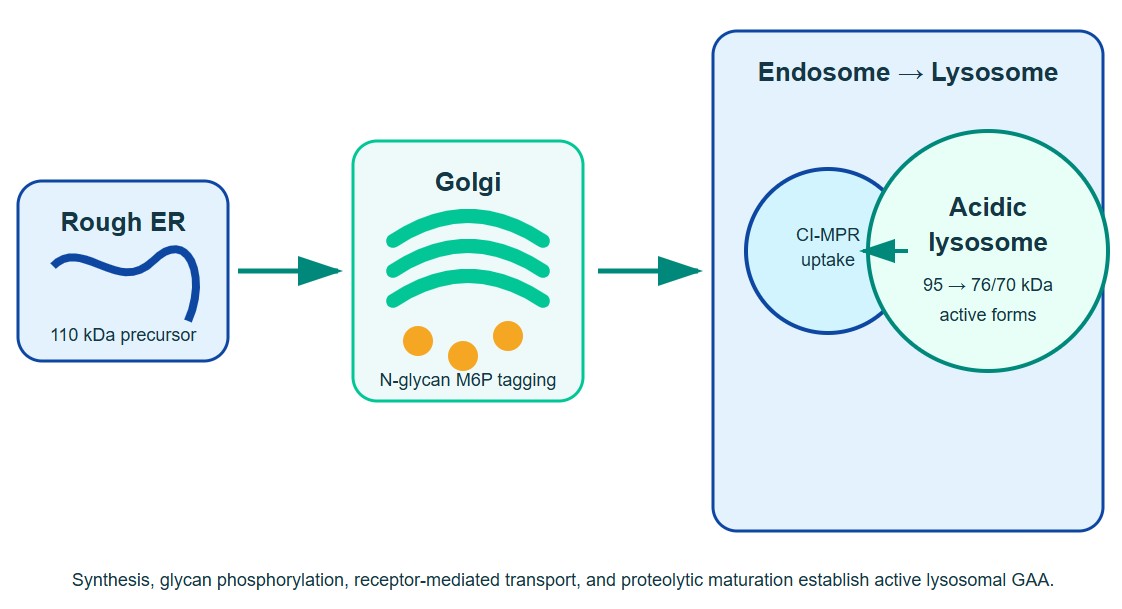
Figure 1: GAA biosynthesis, mannose-6-phosphate-dependent targeting, and proteolytic maturation in the lysosome.
3. Glycogen Breakdown in the Lysosome
Lysosomal glycogen is thought to arise largely through glycophagy, a selective autophagic route that transports glycogen-containing material into lysosomes. The relative contribution of this pathway varies with tissue, developmental stage, nutrient state, and cellular stress. Once glycogen enters the lysosomal lumen, the acidic environment favors GAA activity and allows sequential cleavage from accessible nonreducing ends.
The lysosomal pathway is functionally integrated with autophagy. When GAA is deficient, glycogen-filled lysosomes enlarge and may lose structural integrity. Autophagic material can accumulate, particularly in skeletal muscle fibers, creating regions that interfere with contractile organization and intracellular trafficking. Therefore, measuring total glycogen alone may underestimate the biological effect of GAA deficiency. Lysosomal size, autophagic markers, organelle distribution, and muscle-cell morphology provide complementary information.
In healthy cells, lysosomal glycogen turnover contributes to glucose recycling and organelle homeostasis. In GAA-deficient cells, continued glycogen delivery without adequate hydrolysis produces progressive storage. This imbalance explains why even modest residual activity can strongly influence disease onset and progression: a small difference between substrate influx and degradative capacity accumulates over time.
4. α-1,4 and α-1,6 Glycosidic Bond Hydrolysis
Glycogen consists primarily of α-1,4-linked glucose chains connected by α-1,6 branch points. Effective lysosomal degradation requires cleavage of both linkage types. GAA functions as an exo-acting glucosidase, releasing glucose from nonreducing ends while progressively shortening the polymer. Its activity against α-1,4 bonds supports linear chain digestion, whereas hydrolysis of α-1,6 bonds permits removal of branch structures that would otherwise block continued degradation.
The catalytic mechanism is consistent with a retaining glycosidase reaction in which acidic residues participate in glycosylation and deglycosylation steps. Enzyme activity is strongly pH dependent, and substrate presentation influences apparent kinetics. Small fluorogenic substrates such as 4-methylumbelliferyl-α-D-glucopyranoside are useful for high-throughput activity measurements, but they do not reproduce the branching, size, and steric properties of native glycogen. Assays using glycogen or defined oligosaccharides provide more biologically relevant information about bond specificity and macromolecular substrate turnover.
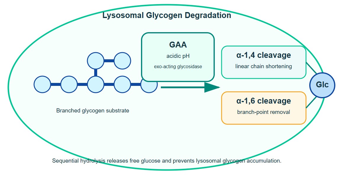
Figure 2: Sequential GAA-mediated hydrolysis of α-1,4 and α-1,6 glycosidic linkages releases glucose from lysosomal glycogen.
5. Tissue Relevance: Skeletal Muscle, Cardiac Muscle, and Respiratory Function
GAA is broadly expressed, but the consequences of deficiency are especially prominent in muscle. High glycogen content, large cell volume, limited regenerative capacity, and sustained mechanical demand make skeletal and cardiac muscle sensitive to lysosomal dysfunction. Tissue response is also shaped by receptor abundance, autophagic state, fiber type, disease stage, and the capacity of extracellular enzyme to cross vascular and interstitial barriers.
| Tissue or System | Biological Dependence | Consequences of GAA Deficiency | Useful Research Readouts |
|---|---|---|---|
| Skeletal muscle | Continuous glycogen turnover, autophagic homeostasis, and contractile integrity | Lysosomal expansion, autophagic buildup, fiber damage, proximal weakness, and reduced mobility | Glycogen content, GAA activity, lysosomal area, LC3/p62 markers, myotube diameter, force generation |
| Cardiac muscle | High energetic demand and tightly organized sarcomeric structure | Marked glycogen storage and hypertrophic cardiomyopathy, especially in severe infantile disease | Cardiomyocyte size, glycogen staining, contractility, calcium handling, ventricular mass in animal models |
| Diaphragm and respiratory muscle | Continuous repetitive contraction and resistance to fatigue | Progressive respiratory muscle weakness and impaired ventilation | Diaphragm force, respiratory mechanics, motor neuron-muscle models, ventilation-related endpoints |
| Motor unit and nervous system | Neuromuscular coordination and axonal support | Potential glycogen accumulation and neural contribution to respiratory or motor dysfunction | Neuromuscular junction morphology, motor neuron models, nerve conduction, tissue GAA distribution |
Cardiac involvement is most prominent in classic infantile-onset disease, where little or no functional GAA is present. Later-onset forms frequently retain some enzyme activity and are dominated by progressive skeletal and respiratory muscle weakness. This spectrum emphasizes that tissue pathology reflects both residual enzyme activity and the duration of substrate accumulation.
6. GAA Deficiency and Glycogen Accumulation
Biallelic pathogenic variants in GAA can reduce enzyme synthesis, folding, catalytic function, intracellular transport, or maturation. The resulting deficiency causes Pompe disease, an autosomal recessive lysosomal storage disorder. Severe variants may produce almost no functional enzyme, whereas other variants permit partial residual activity. In general, lower residual activity is associated with earlier onset and more extensive tissue involvement, although genotype, modifier pathways, immune status, and treatment timing can alter the phenotype.
The pathological sequence begins with lysosomal glycogen retention but develops into a broader disturbance of cellular architecture. Enlarged lysosomes displace myofibrils, membrane rupture can expose glycogen to the cytoplasm, and autophagic debris may accumulate in the center of muscle fibers. These changes can reduce delivery of extracellular recombinant human GAA to affected lysosomes, creating a feedback problem in advanced disease.
GAA deficiency should not be modeled only as increased glycogen. Lysosomal stress, autophagic dysfunction, organelle trafficking, muscle architecture, and residual enzyme activity are interconnected determinants of experimental phenotype.
7. Research Applications of Recombinant Human GAA
Recombinant human GAA is used both as a biochemical reagent and as a model therapeutic enzyme. In cell-free systems, it supports kinetic analysis, substrate-specificity studies, pH profiling, stability testing, glycan characterization, and screening of stabilizers or pharmacological chaperones. In cellular systems, it enables evaluation of M6P receptor-dependent uptake, intracellular trafficking, lysosomal maturation, glycogen clearance, and correction of disease-associated phenotypes.
Alglucosidase alfa and related recombinant formats are also reference tools for comparing next-generation constructs. These may include proteins with altered glycan phosphorylation, receptor-targeting tags, modified half-life, enhanced stability, or alternative production platforms. The most informative comparison depends on the research question: catalytic equivalence requires biochemical normalization, whereas delivery equivalence requires normalization for cellular uptake and lysosomal exposure.
| Research Application | Recommended System | Core Endpoint | Critical Control |
|---|---|---|---|
| Enzyme kinetics | Purified protein with fluorogenic substrate and glycogen | Specific activity, Km, Vmax, pH profile | Substrate blank, heat-inactivated enzyme, matched protein concentration |
| Receptor-mediated uptake | Fibroblasts, myoblasts, myotubes, or receptor-engineered cells | Internalized GAA and intracellular maturation | Excess M6P or receptor-blocking condition |
| Glycogen clearance | GAA-deficient patient cells, iPSC-derived muscle, or animal tissue | Reduction of lysosomal and total glycogen | Untreated disease model and healthy comparator |
| Protein engineering | Side-by-side recombinant variants | Stability, glycan profile, uptake, lysosomal delivery | Activity-normalized and molar-normalized comparisons |
| Immunogenicity research | In vitro immune assays or relevant animal models | Binding antibodies, neutralizing activity, cellular responses | Matched formulation, aggregate assessment, endotoxin control |
| Gene therapy benchmarking | Transduced cells or animal models | Secreted and tissue GAA, cross-correction, glycogen reduction | Vector-only control and recombinant GAA reference |
8. Key Considerations for Experimental Design
8.1 Match the Assay to the Biological Question
Artificial substrates are efficient for screening but can overestimate biological performance. A complete workflow often combines a rapid fluorogenic assay with a glycogen-based assay and a cell-based correction endpoint. For cellular studies, measure not only total GAA activity but also intracellular processing and glycogen reduction.
8.2 Control pH, Temperature, and Incubation Time
GAA is optimized for acidic conditions. Small shifts in assay pH can change activity and apparent kinetic parameters. Buffer identity, ionic strength, substrate concentration, and incubation time should therefore be standardized. Reactions should remain within a linear range for both time and protein concentration.
8.3 Characterize Glycosylation and M6P-Dependent Uptake
Glycan structure affects receptor recognition and trafficking. When comparing recombinant lots or engineered formats, include glycan profiling, M6P-related analysis, receptor-binding assessment, or competitive uptake experiments. CHO-derived and other eukaryotic expression systems may produce different glycan patterns even when the amino-acid sequence is identical.
8.4 Consider Cell Type and Disease Stage
Fibroblasts are convenient for uptake and enzyme assays, but differentiated skeletal muscle models may better reproduce receptor abundance, autophagic buildup, and glycogen storage. Advanced disease models can be less responsive because disrupted trafficking and accumulated autophagic material limit lysosomal access. Dose-response data should therefore be interpreted within the context of baseline pathology.
8.5 Use Orthogonal Readouts
Recommended readouts include GAA activity, Western blotting of precursor and mature forms, total glycogen, lysosome-associated glycogen imaging, LAMP1-positive area, autophagy markers, cell morphology, and functional muscle measurements. Concordance across these endpoints provides stronger evidence than a single activity assay.
8.6 Verify Reagent Quality
Protein purity, aggregation, endotoxin, freeze-thaw history, reconstitution conditions, and storage stability can influence cell-based results. Recombinant enzyme should be aliquoted after reconstitution, handled under conditions that preserve activity, and compared using consistent lot documentation. For immunological studies, trace endotoxin and aggregates are especially important confounders.
9. Summary and Future Perspectives
Acid alpha-glucosidase is the key lysosomal enzyme responsible for converting branched glycogen into glucose through hydrolysis of α-1,4 and α-1,6 glycosidic bonds. Its function depends on a coordinated biological pathway that begins with GAA gene expression and continues through glycoprotein folding, M6P labeling, receptor-dependent trafficking, lysosomal maturation, and substrate access. Deficiency at any of these levels can reduce functional lysosomal activity and promote glycogen storage.
Future GAA research is moving from simple enzyme replacement toward integrated optimization of delivery, intracellular trafficking, glycan design, immune tolerance, autophagic correction, and durable gene-based expression. High-content cell models, patient-derived iPSC muscle systems, organoids, advanced animal models, and quantitative imaging will help connect biochemical enzyme properties with tissue-level correction. At the same time, engineered enzymes with improved receptor engagement or lysosomal delivery may clarify which barriers most strongly limit skeletal-muscle response.
For experimental programs, the central principle is to evaluate lysosomal glycogen degradation as a multistep process rather than as an isolated catalytic reaction. Combining biochemical, cellular, and tissue-level endpoints provides the most reliable framework for studying GAA biology and translating recombinant enzyme findings into Pompe disease research.
The most effective GAA studies connect enzyme quality to receptor uptake, lysosomal maturation, glycogen clearance, and restoration of muscle-cell function.
References
1. Hoefsloot, L. H., et al. (1988). Primary structure and processing of lysosomal alpha-glucosidase; homology with the intestinal sucrase-isomaltase complex. EMBO Journal, 7(6): 1697-1704.
2. Martiniuk, F., et al. (1990). Characterization of the human lysosomal alpha-glucosidase gene. Biochemical Journal, 272(2): 493-497.
3. Wisselaar, H. A., et al. (1993). Human lysosomal alpha-glucosidase: glycosylation, intracellular transport, and maturation. Journal of Biological Chemistry, 268(3): 2223-2231.
4. Moreland, R. J., et al. (2005). Lysosomal acid alpha-glucosidase consists of four different peptides processed from a single-chain precursor. Journal of Biological Chemistry, 280(8): 6780-6791.
5. van der Ploeg, A. T., & Reuser, A. J. J. (2008). Pompe's disease. The Lancet, 372(9646): 1342-1353.
6. Kishnani, P. S., & Steiner, R. D. (2006). Pompe disease diagnosis and management guideline. Genetics in Medicine, 8(5): 267-288.
7. Lim, J. A., et al. (2014). Autophagy in Pompe disease. Methods in Enzymology, 453: 157-184.
8. Raben, N., et al. (2012). Impaired autophagy contributes to muscle atrophy in glycogen storage disease type II. Autophagy, 8(12): 1697-1700.
9. Fukuda, T., et al. (2006). Dysfunction of endocytic and autophagic pathways in a lysosomal storage disease. Annals of Neurology, 59(4): 700-708.
10. van den Hout, J. M. P., et al. (2000). Recombinant human alpha-glucosidase from rabbit milk in Pompe patients. The Lancet, 356(9227): 397-398.
11. Pauly, D. F., et al. (1998). Complete correction of acid alpha-glucosidase deficiency in Pompe disease fibroblasts in vitro, and lysosomally targeted expression in neonatal rat cardiac and skeletal muscle. Gene Therapy, 5(4): 473-480.
12. Leslie, N. D., & Tinkle, B. T. Pompe Disease. GeneReviews®. University of Washington, Seattle.
13. UniProt Consortium. Lysosomal alpha-glucosidase, human GAA (UniProtKB P10253).
14. Zhu, Y., et al. (2009). Glycoengineered acid alpha-glucosidase with improved efficacy at correcting the metabolic aberrations and motor function deficits in a mouse model of Pompe disease. Molecular Therapy, 17(6): 954-963.