
Immunogenicity Considerations for Recombinant Acid Alpha-Glucosidase Research
Protein Attributes, Anti-Drug Antibody Risk, and Integrated Assay Strategies
Abstract
Immunogenicity is a central variable in recombinant enzyme research because an exogenous therapeutic protein can trigger binding antibodies, neutralizing antibodies, infusion-associated reactions, altered pharmacokinetics, or loss of functional response. For recombinant acid alpha-glucosidase (GAA), these risks are especially important in Pompe disease, where treatment depends on repeated systemic exposure, receptor-mediated cellular uptake, lysosomal delivery, and sustained glycogen clearance.
A technically sound immunogenicity program should not rely on a single antibody assay. Instead, it should combine product characterization, tiered anti-drug antibody testing, functional neutralization assays, exposure measurements, enzyme activity, cellular uptake, and disease-relevant outcome data. This review summarizes the principal risk factors, assay approaches, and interpretation strategies for GAA immunogenicity research.
GAA immunogenicity, recombinant enzyme immunogenicity, alglucosidase alfa antibodies, Pompe disease ERT immune response, therapeutic protein immunogenicity
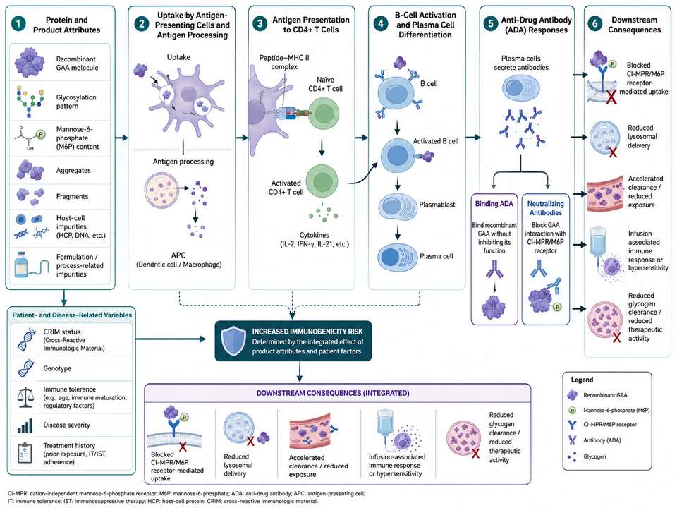
Figure 1: Integrated immunogenicity pathway for recombinant GAA, from protein attributes and antigen presentation to binding antibodies, neutralizing activity, altered exposure, and reduced lysosomal correction.
1. Why Immunogenicity Matters in Enzyme Therapy Research
Enzyme replacement research requires repeated administration of a relatively large protein dose. This creates multiple opportunities for immune recognition, particularly when endogenous expression is absent or very low, when the recombinant protein contains structural or glycan differences, or when product-related impurities promote innate immune activation. Antibody formation may remain clinically silent, but in some settings it can modify drug exposure, block receptor binding, inhibit catalytic activity, or contribute to hypersensitivity and immune-complex effects.
| Potential Immune Outcome | Mechanism | Research Consequence |
|---|---|---|
| Binding ADA | Antibodies bind one or more GAA epitopes without measurable functional inhibition | May change clearance or complicate pharmacokinetic interpretation |
| Neutralizing ADA | Antibodies block enzyme activity, receptor binding, uptake, or intracellular function | Can reduce lysosomal delivery and glycogen clearance |
| Infusion-associated response | Cytokine release, complement activation, or immediate hypersensitivity | May affect tolerability and dosing continuity |
| Immune-complex effects | Persistent antigen-antibody complexes deposit or activate inflammatory pathways | May create organ-specific safety signals |
2. Recombinant GAA as a Therapeutic Protein
Recombinant GAA is a glycosylated lysosomal hydrolase whose biological function depends on more than catalytic activity alone. The molecule must remain folded, retain mannose-6-phosphate-containing glycans, interact with cellular receptors, traffic through endosomes, and mature in lysosomes. Changes in sequence, glycosylation, oxidation, deamidation, aggregation, fragmentation, or formulation can therefore influence both potency and immunogenic potential.
| Product Attribute | Functional Importance | Immunogenicity Relevance |
|---|---|---|
| Primary sequence | Defines catalytic and structural domains | Non-native variants may create new T-cell or B-cell epitopes |
| Glycosylation | Supports receptor-mediated uptake and intracellular routing | Unusual glycans may alter clearance or immune recognition |
| Higher-order structure | Maintains enzyme activity and stability | Partially unfolded protein may expose cryptic epitopes |
| Aggregation state | Affects dose uniformity and uptake | Multivalent aggregates can enhance antigen presentation |
| Process impurities | Should be minimized for research consistency | Host-cell proteins, DNA, endotoxin, or particulates may act as immune adjuvants |
3. Anti-Drug Antibody Responses
Anti-drug antibody testing is commonly organized as a tiered process. Samples are first screened for potential reactivity, then confirmed for specificity, and finally characterized for titer, isotype, persistence, epitope profile, and neutralizing function when appropriate. A positive binding result should not automatically be interpreted as clinically or biologically meaningful.
| Assay Tier | Purpose | Typical Output |
|---|---|---|
| Screening assay | Detect potentially reactive samples with high sensitivity | Screen-positive or screen-negative |
| Confirmatory assay | Demonstrate GAA-specific inhibition or competition | Confirmed ADA-positive or negative |
| Titer assay | Estimate relative antibody magnitude | Endpoint dilution or relative titer |
| Characterization assay | Define isotype, persistence, epitope, or neutralizing activity | Functional and mechanistic interpretation |
ADA positivity, ADA titer, and neutralizing activity are related but not interchangeable measurements. Their significance should be assessed together with exposure, enzyme activity, uptake, safety, and disease-response data.
4. Factors Influencing Immunogenicity
Immunogenicity is multifactorial. Product quality, treatment regimen, route of administration, dose frequency, immune status, genotype, residual endogenous protein, concurrent inflammation, and prior exposure can all change the probability and consequences of an immune response.
| Risk Category | Examples | Suggested Assessment |
|---|---|---|
| Product related | Aggregation, oxidation, clipping, non-native glycans, impurities | Orthogonal analytical characterization |
| Process related | Host-cell proteins, residual DNA, endotoxin, leachables | Impurity profiling and lot comparability |
| Treatment related | Dose, schedule, infusion rate, treatment interruption | Exposure-response and longitudinal ADA analysis |
| Patient related | CRIM status, genotype, immune maturity, HLA background | Prospective stratification and covariate analysis |
| Disease related | Inflammation, organ damage, baseline severity | Clinical and biomarker context |
5. Protein Aggregation and Impurity Risks
Aggregates and certain process impurities are among the most actionable product-related risk factors. Aggregates can increase apparent valency, promote uptake by antigen-presenting cells, expose non-native surfaces, and complicate dose calculations. Even low levels of endotoxin or host-cell proteins can confound cytokine or immune-cell assays by creating adjuvant-like stimulation unrelated to GAA itself.
| Risk Attribute | Recommended Method | Interpretive Value |
|---|---|---|
| Soluble aggregates | SEC-HPLC, analytical ultracentrifugation | Quantifies dimers and higher-order species |
| Subvisible particles | Light obscuration, micro-flow imaging | Detects particles that may stimulate innate immunity |
| Structural instability | DSC, DLS, intrinsic fluorescence, stress studies | Identifies conditions that expose non-native conformations |
| Host-cell proteins | HCP ELISA and LC-MS | Assesses residual process-related antigens |
| Endotoxin | LAL or recombinant factor C assay | Helps distinguish product immunogenicity from innate activation |
6. Patient-Related and Disease-Related Variables
In Pompe disease, the presence or absence of endogenous cross-reactive immunologic material is a major immunological variable. Patients with little or no native GAA may have reduced immune tolerance to administered enzyme. Age, genotype, residual activity, previous treatment, immune-modulating medication, infection, and baseline disease severity can further influence antibody development and clinical relevance.
- CRIM status: absence of endogenous GAA protein can increase the risk of strong antibody responses.
- Genotype and residual expression: may affect immune tolerance and baseline disease biology.
- Age at first exposure: immune maturity and disease urgency can shape response.
- Prior ERT exposure: existing antibodies may interfere with comparative studies or switching experiments.
- Inflammatory state: infection, tissue damage, or concurrent immune activation may increase antigen presentation.
7. In Vitro and Preclinical Immunogenicity Assays
No single preclinical assay predicts human immunogenicity with certainty. The most informative programs combine analytical risk assessment with immune-cell assays, animal studies, and functional neutralization testing. Assays should be selected according to the hypothesized mechanism rather than used as a generic checklist.
| Assay | Question Addressed | Key Limitation |
|---|---|---|
| T-cell proliferation or cytokine assay | Can GAA-derived peptides activate donor T cells? | Donor variability and uncertain clinical translation |
| Dendritic-cell uptake and activation | Does a formulation or aggregate increase antigen presentation? | Highly sensitive to endotoxin and impurities |
| Binding ADA assay | Are GAA-reactive antibodies present? | Drug interference and matrix effects |
| Enzyme neutralization assay | Do antibodies inhibit catalytic activity? | May miss uptake-blocking antibodies |
| Cell-based uptake neutralization assay | Do antibodies block receptor binding, endocytosis, or lysosomal delivery? | Requires robust, disease-relevant cell models |
| GAA-knockout animal study | Does repeated dosing produce antibodies and altered efficacy? | Species-specific immune biology may not predict humans |
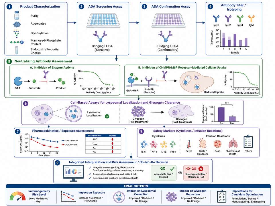
Figure 2: Tiered workflow linking product characterization, ADA screening and confirmation, neutralization assays, cellular uptake, pharmacokinetics, safety markers, and glycogen-clearance readouts.
8. Interpreting Antibody and Activity Data
Longitudinal interpretation is essential. A transient low-titer ADA signal may have little functional effect, while persistent high-titer antibodies accompanied by reduced circulating GAA, impaired cellular uptake, lower tissue enzyme activity, or diminished glycogen clearance are more concerning. Timing relative to dosing is also critical because circulating drug can mask ADA detection.
| Observed Pattern | Possible Interpretation | Follow-Up |
|---|---|---|
| ADA positive, normal exposure and activity | Binding antibodies without clear functional effect | Continue longitudinal monitoring |
| ADA titer rises, exposure declines | Antibody-mediated clearance may be occurring | Assess PK, immune complexes, and neutralization |
| Normal plasma exposure, reduced cell uptake | Receptor-blocking or uptake-neutralizing antibodies | Use a cell-based uptake assay |
| Normal uptake, reduced enzyme activity | Catalytic neutralization or intracellular processing issue | Measure activity and lysosomal maturation |
| Antibody signal plus infusion reactions | Potential immune-mediated safety response | Correlate with complement, tryptase, and cytokines |
9. Risk Reduction Strategies in Research Development
Risk reduction begins with molecule and process design. Maintaining native-like structure, controlling aggregates, minimizing impurities, optimizing formulation, and using consistent storage conditions can reduce avoidable immune stimuli. Study design should include baseline samples, appropriate positive controls, drug-tolerance assessment, matched sampling times, and predefined criteria for functional follow-up.
| Development Stage | Risk-Reduction Action | Expected Benefit |
|---|---|---|
| Molecule design | Minimize non-native sequence changes and unstable domains | Reduces novel epitope and unfolding risk |
| Process development | Control aggregates, HCP, DNA, endotoxin, and particulates | Reduces adjuvant-like stimulation |
| Formulation | Optimize pH, excipients, freeze-thaw, and agitation tolerance | Maintains structural integrity during handling |
| Assay development | Validate sensitivity, specificity, drug tolerance, and cut points | Improves reliability of ADA conclusions |
| Preclinical design | Collect exposure, ADA, neutralization, tissue activity, and pathology data together | Supports integrated mechanistic interpretation |
Use the same well-characterized GAA reference material across binding, activity, uptake, and neutralization assays whenever possible. This improves comparability between analytical and functional datasets.
10. Summary
Immunogenicity assessment for recombinant GAA is not simply an antibody-detection exercise. It is an integrated evaluation of molecule quality, immune recognition, exposure, receptor-mediated uptake, lysosomal activity, glycogen clearance, and safety. The most informative studies distinguish binding antibodies from neutralizing antibodies and identify whether functional interference occurs at the level of circulation, receptor engagement, internalization, intracellular trafficking, or catalysis.
For GAA research programs, robust conclusions require orthogonal assays, longitudinal sampling, appropriate controls, and explicit consideration of product-, patient-, and disease-related risk factors. This framework supports more reliable comparison of alglucosidase alfa and recombinant GAA candidates during enzyme replacement and next-generation therapeutic development.
References
1. U.S. Food and Drug Administration. LUMIZYME (alglucosidase alfa) prescribing information, 2024.
2. European Medicines Agency. Guideline on Immunogenicity Assessment of Therapeutic Proteins, Revision 1.
3. Banugaria, S. G., et al. (2011). The impact of antibodies on clinical outcomes in diseases treated with therapeutic protein: lessons learned from infantile Pompe disease. Genet Med, 13(8): 729-736.
4. Kishnani, P. S., et al. (2010). Cross-reactive immunologic material status affects treatment outcomes in Pompe disease infants. Mol Genet Metab, 99(1): 26-33.
5. Patel, T. T., et al. (2012). The impact of antibodies in late-onset Pompe disease: a case series and literature review. Mol Genet Metab, 106(3): 301-309.
6. Desai, A. K., et al. (2024). Immunophenotype associated with high sustained antibody titers against enzyme replacement therapy in infantile-onset Pompe disease. Front Immunol, 14: 1296911.
7. Doerfler, P. A., et al. (2016). Immune tolerance and gene therapy approaches in Pompe disease. Mol Ther Methods Clin Dev, 3: 16046.
8. Rosenberg, A. S., & Sauna, Z. E. (2018). Immunogenicity assessment during the development of protein therapeutics. J Pharm Pharmacol, 70(5): 584-594.
9. van Beers, M. M. C., & Bardor, M. (2012). Minimizing immunogenicity of biopharmaceuticals by controlling critical quality attributes. Biotechnol J, 7(12): 1473-1484.
10. Moussa, E. M., et al. (2016). Immunogenicity of therapeutic protein aggregates. J Pharm Sci, 105(2): 417-430.