
Cell-Based Evaluation of Recombinant Acid Alpha-Glucosidase Uptake and Lysosomal Function
An Experimental Guide to Cellular Uptake, Lysosomal Delivery, and Functional Glycogen Clearance
Abstract
Cell-based testing provides a functional bridge between biochemical characterization of recombinant acid alpha-glucosidase and its expected intracellular activity. A useful assay should determine whether the enzyme binds the cell surface, enters the endocytic pathway, reaches lysosomes, undergoes intracellular processing, restores acid alpha-glucosidase activity, and reduces accumulated glycogen. These endpoints are especially relevant when comparing recombinant enzyme preparations, glycoengineered variants, formulations, or uptake-enhancing strategies in Pompe disease research models.
This guide outlines practical approaches for evaluating recombinant acid alpha-glucosidase uptake and lysosomal function using complementary imaging, biochemical, and molecular methods. Emphasis is placed on experimental design, receptor-dependent uptake, assay controls, normalization, and interpretation of glycogen clearance data.
GAA cell assay, recombinant GAA uptake, Pompe disease cell model, lysosomal enzyme uptake, mannose-6-phosphate receptor, glycogen clearance assay, acid alpha-glucosidase protein
1. Why Cell-Based GAA Testing Matters
Purified-enzyme activity assays confirm catalytic potential under defined buffer conditions, but they do not establish whether a recombinant enzyme can function inside a relevant cell. For lysosomal enzymes, biological performance depends on several linked events: receptor recognition, endocytosis, endosomal trafficking, lysosomal delivery, proteolytic maturation, and substrate access. A preparation with strong activity against a soluble fluorogenic substrate may still show limited cellular efficacy if uptake is inefficient or intracellular routing is incomplete.
Cell-based assays therefore add translational context to the characterization of recombinant GAA. They can support lot comparison, formulation screening, glycan-dependent uptake studies, mechanism-of-action research, and selection of concentrations for downstream animal studies.
| Question | Recommended Readout | Interpretive Value |
|---|---|---|
| Does GAA bind and enter cells? | Cell-associated fluorescence, flow cytometry, or labeled-enzyme uptake | Measures initial uptake but not necessarily lysosomal delivery |
| Does internalized GAA reach lysosomes? | Co-localization with LAMP1/LAMP2 or LysoTracker | Confirms intracellular routing to the target compartment |
| Is intracellular enzyme active? | GAA activity in washed cell lysates | Demonstrates functional activity after uptake |
| Does treatment correct the disease phenotype? | Glycogen reduction and lysosomal phenotype improvement | Provides the most disease-relevant functional endpoint |
2. Common Cell Models for Pompe Disease Research
Model selection should match the experimental question. High-throughput uptake screening may favor robust immortalized cells, whereas disease-relevant functional testing benefits from patient-derived or genetically defined GAA-deficient cells. Skeletal muscle lineage models are particularly useful because muscle pathology is a major feature of Pompe disease, but fibroblasts and engineered cell lines often provide more reproducible starting systems.
| Cell Model | Advantages | Typical Applications | Key Limitations |
|---|---|---|---|
| Patient-derived fibroblasts | Endogenous disease genotype; accessible and relatively easy to culture | Uptake, lysosomal localization, enzyme recovery, glycogen measurement | Donor variability; limited representation of skeletal muscle pathology |
| Primary or immortalized myoblasts | Relevant muscle lineage and differentiation capacity | Myotube uptake, lysosomal correction, glycogen clearance | Differentiation state and passage number can affect results |
| GAA-knockout engineered cells | Defined genetic background and strong negative baseline | Assay development, concentration-response analysis, mechanism studies | May not reproduce all patient-specific phenotypes |
| iPSC-derived myocytes | Patient-specific genotype with disease-relevant differentiation | Mechanistic studies, long-term correction, phenotypic rescue | Cost, complexity, maturation variability, and batch effects |
| Wild-type receptor-competent cell lines | Robust growth and convenient screening | Receptor dependence and comparative uptake studies | Low disease relevance and possible endogenous GAA background |
Use a simple, reproducible model for assay optimization and a disease-relevant model for confirmation. Consistent conclusions across two cell systems are stronger than extensive optimization in only one model.
3. Recombinant GAA Uptake Mechanisms
Extracellular acid alpha-glucosidase can be internalized through receptor-mediated endocytosis. Product glycosylation, mannose-6-phosphate content, receptor abundance, cell differentiation state, enzyme concentration, exposure time, and competition from endogenous ligands may all influence uptake efficiency.
A common experimental sequence includes treatment with recombinant enzyme, removal of extracellular material by repeated washing, optional surface stripping, recovery in enzyme-free medium, and analysis of intracellular localization or activity. Uptake should be distinguished from nonspecific adsorption by using low-temperature binding controls, receptor competition, or acid wash conditions where appropriate.
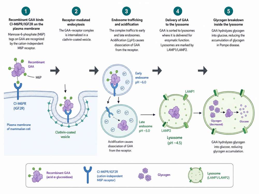
Figure 1: Recombinant GAA uptake and lysosomal delivery. The enzyme binds a cell-surface mannose-6-phosphate receptor, enters the endocytic pathway, traffics through endosomes, reaches lysosomes, and contributes to glycogen degradation.
4. Mannose-6-Phosphate Receptor-Mediated Delivery
The cation-independent mannose-6-phosphate receptor is a major route for cellular uptake of phosphorylated lysosomal enzymes. Mannose-6-phosphate-bearing glycans on recombinant GAA are recognized at the plasma membrane, followed by receptor-ligand internalization and trafficking through endosomal compartments. Acidification promotes ligand dissociation, while the receptor is recycled and the enzyme proceeds toward lysosomes.
| Experimental Approach | Expected Observation | Interpretation |
|---|---|---|
| Excess free mannose-6-phosphate | Reduced GAA uptake or intracellular activity | Supports M6P receptor-dependent internalization |
| Receptor-blocking antibody | Reduced cell-associated GAA signal | Provides orthogonal evidence for receptor involvement |
| 4°C binding condition | Surface binding with limited internalization | Separates binding from active endocytosis |
| Receptor-low versus receptor-high cells | Different uptake efficiency | Links cell phenotype to enzyme internalization capacity |
Competition controls must be optimized carefully. Excess competitor can alter medium osmolarity or affect other pathways, while receptor-blocking antibodies may vary in affinity and epitope accessibility. A concentration series is preferable to a single inhibitory condition.
5. Lysosomal Localization Assessment
Internalization alone is not sufficient evidence of lysosomal delivery. Fluorescence microscopy can determine whether GAA signal overlaps with lysosomal markers such as LAMP1, LAMP2, or LysoTracker. Confocal imaging is generally preferred over widefield imaging because optical sectioning reduces apparent overlap from signals located above or below the same focal plane.
| Method | Recommended Output | Important Controls |
|---|---|---|
| Immunofluorescence | GAA/LAMP1 merged images and Pearson or Manders coefficients | Secondary-only control, untreated cells, single-stain controls |
| Fluorescently labeled GAA | Time-resolved internalization and lysosomal overlap | Verify that labeling does not impair uptake or activity |
| Subcellular fractionation | GAA activity or protein enrichment in lysosome-associated fractions | Fraction purity markers and recovery assessment |
| Immunoelectron microscopy | Ultrastructural localization within lysosomal compartments | Specific labeling and adequate sampling |
Quantitative image analysis should be performed with fixed acquisition settings, blinded fields, and predefined segmentation rules. Reporting only a representative image can overstate biological consistency.
6. Glycogen Clearance Readouts
Reduction of intracellular glycogen is a central functional endpoint for a Pompe disease cell model. The most informative designs measure baseline glycogen accumulation, apply recombinant GAA under defined exposure conditions, allow sufficient time for lysosomal processing, and quantify residual glycogen using a method compatible with the expected concentration range.
| Method | Strengths | Limitations | Best Use |
|---|---|---|---|
| Enzymatic glucose-release assay | Quantitative, scalable, and compatible with plate formats | Requires careful background correction and normalization | Routine concentration-response studies |
| PAS staining | Shows cell-level and spatial glycogen distribution | Semi-quantitative and sensitive to staining conditions | Phenotypic visualization and morphology |
| Fluorometric glycogen assay | High sensitivity with relatively low sample volume | Potential interference from sample matrix | Low-abundance samples and screening |
| Electron microscopy | Visualizes lysosomal glycogen and ultrastructural correction | Low throughput and labor intensive | Mechanistic confirmation |
Results should be normalized to total protein, DNA, cell number, or another validated biomass metric. Apparent glycogen reduction caused by cytotoxicity or cell loss should not be interpreted as functional correction.
7. Enzyme Activity Recovery in Cells
Intracellular activity assays help determine whether internalized GAA remains catalytically competent. A common method uses 4-methylumbelliferyl-alpha-D-glucopyranoside under acidic conditions, with fluorescence measured after stopping the reaction. Cell monolayers should be washed thoroughly before lysis to minimize carryover of extracellular enzyme.
Recommended Workflow
- Treat cells with a concentration series of recombinant GAA for a defined uptake period.
- Wash repeatedly with cold buffer; use a validated surface-removal step when needed.
- Lyse cells in a buffer compatible with the activity assay and protein quantification.
- Measure GAA activity at acidic pH with substrate blanks and calibration standards.
- Normalize activity to total protein, DNA, or cell number.
- Compare activity recovery with glycogen clearance to confirm functional linkage.
A high intracellular fluorescence signal with limited enzyme activity may indicate label accumulation, inactive protein, incomplete lysosomal processing, or assay interference. Pair uptake measurements with a catalytic readout.
8. Imaging, Western Blot, and Biochemical Methods
No single analytical method fully describes uptake and function. An orthogonal strategy combines spatial localization, protein processing, catalytic activity, and substrate reduction. The selected method panel should reflect the maturity of the study and the amount of available sample.
| Technique | Primary Question | Typical Output | Common Pitfall |
|---|---|---|---|
| Confocal microscopy | Where is GAA located? | Co-localization with lysosomal markers | Overinterpretation of visually merged signals |
| Flow cytometry | How many cells take up GAA? | Positive-cell percentage and signal intensity | Failure to distinguish surface-bound from internalized enzyme |
| Western blot | Is GAA present and processed? | Precursor and mature GAA-associated bands | Antibody specificity and unequal loading |
| Cell lysate activity assay | Is internalized GAA active? | Activity per unit protein or per cell | Residual extracellular enzyme contamination |
| Glycogen assay | Does treatment correct substrate accumulation? | Residual glycogen relative to control | Cell loss masquerading as glycogen reduction |
| qPCR or transcript profiling | Does treatment alter lysosomal stress pathways? | Expression of lysosomal and autophagy-related genes | Transcript changes do not prove functional correction |
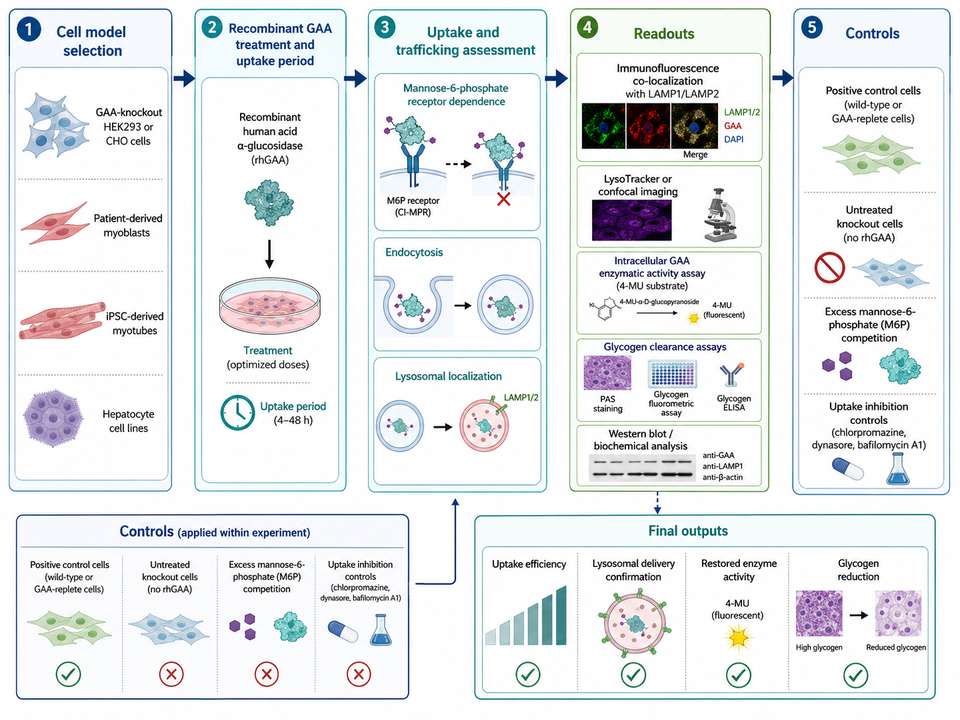
Figure 2: Integrated cell-based GAA evaluation workflow combining uptake, lysosomal localization, protein processing, enzyme activity recovery, and glycogen clearance.
9. Positive and Negative Controls
Controls should test assay performance, biological specificity, and data interpretation. At minimum, each experiment should include untreated disease cells, a positive enzyme-treatment condition, and a control that challenges receptor-mediated uptake. Additional controls may be necessary for fluorescent labeling, antibody specificity, cytotoxicity, and normalization.
| Control Type | Example | Purpose |
|---|---|---|
| Positive biological control | Wild-type cells or a previously qualified recombinant GAA preparation | Defines expected activity or correction |
| Negative disease control | Untreated GAA-deficient cells | Defines baseline enzyme activity and glycogen burden |
| Inactive enzyme control | Heat-inactivated or catalytically inactive GAA | Distinguishes uptake from catalytic correction |
| Receptor competition control | Excess mannose-6-phosphate or receptor-blocking reagent | Tests receptor-mediated uptake |
| Surface-binding control | 4°C incubation or surface stripping | Separates membrane association from internalization |
| Imaging control | Secondary-only and single-color samples | Assesses nonspecific signal and spectral overlap |
| Viability control | Cell viability or total-cell measurement | Excludes cytotoxicity-driven false improvement |
10. Experimental Limitations and Best Practices
| Experimental Issue | Potential Impact | Best Practice |
|---|---|---|
| Variable receptor expression | Changes apparent uptake between cell types or passages | Measure or qualify receptor expression and control passage number |
| Differences in GAA glycosylation | Alter receptor binding and cellular delivery | Compare preparations using matched protein mass and activity inputs |
| Extracellular carryover | Inflates intracellular activity measurements | Use stringent washing and validate surface-removal procedures |
| Fluorescent-label interference | Changes uptake, trafficking, or catalytic activity | Test labeled versus unlabeled enzyme in parallel |
| Assay saturation | Hides differences between enzyme preparations | Establish linear ranges for concentration, time, and detector response |
| Cell loss or toxicity | Creates false glycogen reduction | Include viability and biomass normalization in every experiment |
| Single-endpoint interpretation | Cannot distinguish uptake from functional correction | Combine localization, activity, and glycogen readouts |
| Donor or differentiation variability | Reduces reproducibility | Use biological replicates, standardized differentiation, and predefined acceptance criteria |
Practical Best-Practice Checklist
- Define whether the primary objective is uptake, lysosomal delivery, catalytic recovery, or phenotypic correction.
- Use concentrations that cover sub-saturating through near-maximal response rather than a single dose.
- Confirm that enzyme exposure time and post-treatment chase time are appropriate for the selected endpoint.
- Use at least one receptor-specific control and one disease-relevant functional endpoint.
- Report normalization procedures, cell passage, differentiation state, imaging settings, and replicate structure.
- Validate major findings in a second cell model whenever possible.
A robust cell-based GAA assay should connect receptor-dependent uptake with lysosomal localization, recovered catalytic activity, and measurable glycogen reduction. Agreement across these endpoints provides stronger evidence of functional intracellular delivery than any single assay alone.
References
1. Reuser, A. J. J., et al. (2008). Enzyme therapy for Pompe disease: from science to industrial enterprise. Eur J Pediatr, 167(4): 385-394.
2. van der Ploeg, A. T., & Reuser, A. J. J. (2008). Pompe's disease. Lancet, 372(9646): 1342-1353.
3. Kishnani, P. S., et al. (2007). Recombinant human acid alpha-glucosidase: major clinical benefits in infantile-onset Pompe disease. Neurology, 68(2): 99-109.
4. Zhu, Y., et al. (2009). Glycoengineered acid alpha-glucosidase with improved efficacy at correcting the metabolic aberrations and motor function deficits in a mouse model of Pompe disease. Mol Ther, 17(6): 954-963.
5. Koeberl, D. D., Kishnani, P. S., & Chen, Y. T. (2007). Glycogen storage disease types I and II: treatment updates. J Inherit Metab Dis, 30(2): 159-164.
6. Cardone, M., et al. (2008). The role of autophagy in the pathogenesis of Pompe disease. Cell Death Differ, 15(9): 1470-1478.
7. Raben, N., et al. (2003). Enzyme replacement therapy in the mouse model of Pompe disease. Mol Ther, 8(4): 576-584.
8. Lim, J. A., et al. (2014). Pompe disease: from pathophysiology to therapy and back again. Front Aging Neurosci, 6: 177.
9. Fukuda, T., et al. (2006). Dysfunction of endocytic and autophagic pathways in a lysosomal storage disease. Ann Neurol, 59(4): 700-708.
10. Meikle, P. J., et al. (2006). Diagnosis of lysosomal storage disorders: evaluation of lysosome-associated membrane protein LAMP-1 as a diagnostic marker. Clin Chem, 52(1): 56-64.