
Analytical Characterization Strategies for Recombinant Acid Alpha-Glucosidase
Identity, Purity, Activity, Glycosylation, Aggregation, and Impurity Control
Abstract
Recombinant acid alpha-glucosidase (GAA) is a complex lysosomal glycoprotein whose research performance depends on more than nominal protein concentration. Identity, structural integrity, catalytic activity, glycosylation, mannose-6-phosphate-related receptor binding, aggregation, host-cell impurities, and endotoxin can each influence experimental outcomes. A fit-for-purpose characterization strategy therefore combines orthogonal physicochemical, biochemical, and cell-based methods rather than relying on a single purity or activity result.
This technical article reviews analytical approaches for recombinant Acid alpha-glucosidase used in Pompe disease research. The recommended test panel should be matched to the intended use: routine biochemical assays may require a focused identity–purity–activity package, whereas receptor-mediated uptake, glycogen-clearance, formulation, or comparability studies require deeper analysis of glycosylation, aggregation, and biological function.
recombinant GAA characterization, acid alpha-glucosidase purity, GAA glycosylation analysis, SDS-PAGE, HPLC, enzyme activity assay, alglucosidase alfa QC
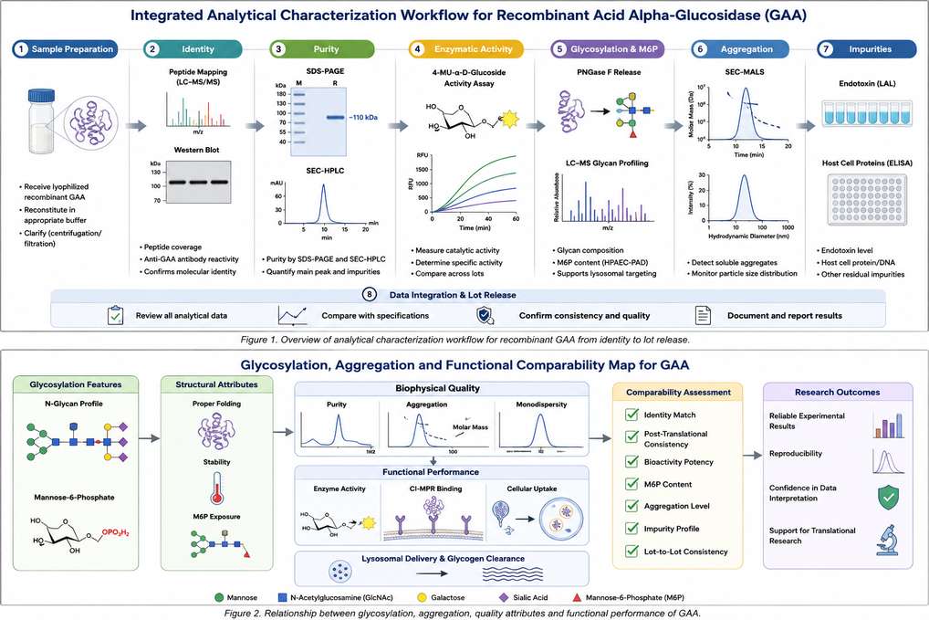
Figure 1: Orthogonal characterization workflow linking identity, purity, aggregation, catalytic activity, glycosylation, mannose-6-phosphate-related uptake, and impurity testing.
1. Characterization Goals for Recombinant GAA
The first step is to define which product attributes are critical for the planned experiment. For a recombinant lysosomal enzyme, analytical quality is multidimensional: the protein must be the correct molecule, remain sufficiently intact and monomeric, retain catalytic competence, and preserve the glycan features needed for receptor-mediated lysosomal delivery.
| Quality Attribute | Key Question | Typical Research Impact |
|---|---|---|
| Identity | Is the sample the intended human GAA protein? | Prevents use of mislabeled, truncated, or cross-contaminated material |
| Purity and integrity | Are major fragments, contaminating proteins, or abnormal bands present? | Affects concentration assignment, assay specificity, and reproducibility |
| Catalytic activity | Does the enzyme hydrolyze a suitable substrate under defined conditions? | Confirms biochemical function and supports specific-activity calculation |
| Glycosylation and M6P-related attributes | Are receptor-relevant glycans present and accessible? | Influences CI-MPR binding, cellular uptake, and lysosomal delivery |
| Aggregation | What proportion is monomeric versus oligomeric or particulate? | Can alter uptake, apparent potency, stability, and immunogenicity risk |
| Process impurities | Are endotoxin, host-cell proteins, DNA, or residual reagents controlled? | Reduces assay interference and nonspecific cellular responses |
Specifications used for release testing are not identical to a full characterization package. Research programs should select methods that are sensitive to the attributes most likely to affect the intended assay or biological conclusion.
2. Identity Testing
Identity should be established using at least one sequence- or epitope-specific method. For higher-confidence studies, an orthogonal combination is preferred because intact mass, peptide-level sequence coverage, and antibody recognition answer different questions.
| Method | Information Provided | Strengths | Limitations |
|---|---|---|---|
| Peptide mapping by LC-MS/MS | Sequence coverage and peptide-level confirmation | High specificity; can identify modifications and clipping sites | Requires specialized instrumentation and data analysis |
| Intact-mass LC-MS | Overall molecular-mass distribution | Rapid confirmation of gross consistency | Glycan heterogeneity broadens the mass envelope |
| Western blot | Recognition by anti-GAA antibody | Accessible and useful for routine confirmation | Antibody cross-reactivity may limit specificity |
| N-terminal sequencing | N-terminal identity and processing | Useful for checking maturation or truncation | Blocked or heterogeneous termini can complicate analysis |
| ELISA or immunoassay | GAA-specific antigen content | High throughput and quantitative | Measures epitope recognition rather than full molecular identity |
3. SDS-PAGE and HPLC Purity Analysis
SDS-PAGE and chromatographic purity methods should be interpreted together. Electrophoresis provides a visual profile of major protein species, while HPLC offers higher-resolution quantification of size- or charge-related heterogeneity.
3.1 SDS-PAGE
Reducing and non-reducing SDS-PAGE can reveal major degradation products, disulfide-linked species, and shifts in apparent molecular weight associated with glycosylation. Densitometry is useful for trend analysis, but co-migrating impurities and staining nonlinearity can limit absolute purity estimates.
3.2 HPLC and Electrophoretic Methods
| Method | Main Attribute | Recommended Interpretation |
|---|---|---|
| SEC-HPLC / SEC-UPLC | Monomer, soluble aggregates, and fragments | Primary quantitative method for size variants; monitor recovery and nonspecific column interactions |
| Ion-exchange HPLC | Charge heterogeneity | Useful for deamidation, sialylation-related shifts, and process comparability |
| Reverse-phase HPLC | Hydrophobic variants and selected impurities | Can detect oxidation or clipping but may denature the protein |
| Capillary electrophoresis-SDS | Size purity and fragments | Offers improved automation and quantitative precision over slab gels |
| Imaged capillary isoelectric focusing | Isoelectric-point distribution | Supports charge-profile comparison among lots or formulations |
4. Enzymatic Activity Testing
Biochemical activity assays confirm substrate hydrolysis but must be designed carefully. GAA activity is sensitive to pH, temperature, substrate type, incubation time, and enzyme concentration. A fluorogenic substrate such as 4-methylumbelliferyl-α-D-glucopyranoside is convenient for routine testing, whereas glycogen-based assays more closely reflect the natural substrate.
| Assay Format | Typical Readout | Best Use | Critical Controls |
|---|---|---|---|
| 4-MU fluorogenic assay | Fluorescence from released 4-methylumbelliferone | Routine activity, kinetics, stability, and lot comparison | Blank, no-enzyme control, reference GAA, linearity range |
| Glycogen hydrolysis assay | Glucose or reducing-sugar release | Natural-substrate confirmation | Substrate blank, glucose standard curve, time-course linearity |
| Cell-lysate activity recovery | Intracellular GAA activity after treatment | Integrated uptake and lysosomal delivery studies | Untreated cells, uptake competition, normalization to protein or cell number |
| Glycogen-clearance assay | Reduced cellular glycogen by PAS, fluorescence, or biochemical quantification | Functional correction in Pompe disease models | GAA-deficient negative control and corrected positive control |
Specific activity should be reported with complete assay conditions and the concentration method used. Apparent activity may change if concentration is assigned by UV absorbance, total-protein assays, or nominal vial content.
5. Glycosylation and Mannose-6-Phosphate-Related Analysis
Human lysosomal GAA is heavily N-glycosylated, and mannose-6-phosphate (M6P) residues support binding to the cation-independent mannose-6-phosphate receptor. Therefore, glycan characterization is especially important when the research objective involves muscle-cell uptake, lysosomal targeting, or comparison of expression platforms.
| Analytical Approach | Attribute Assessed | Application |
|---|---|---|
| Released N-glycan profiling by HILIC-FLD or LC-MS | Relative distribution of high-mannose, hybrid, complex, and sialylated glycans | Lot comparison and expression-system evaluation |
| Glycopeptide LC-MS/MS | Site-specific glycan occupancy and microheterogeneity | Detailed structure–function investigation |
| Monosaccharide or sialic-acid analysis | Global glycan composition | Supportive characterization and comparability |
| M6P quantification | Total or accessible phosphomannose-related content | Screening for receptor-targeting potential |
| CI-MPR binding assay | Functional receptor interaction | Bridges glycan structure to uptake-relevant binding |
| Cellular uptake with excess M6P competition | Receptor-dependent internalization | Confirms that uptake is mediated substantially through M6P receptors |
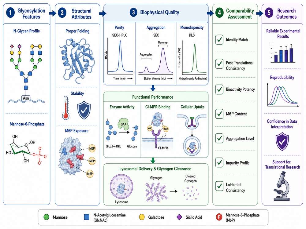
Figure 2: Structure–function map connecting GAA glycosylation, M6P-related receptor binding, aggregation state, catalytic activity, cellular uptake, and glycogen clearance.
6. Aggregation Assessment
Aggregation should be evaluated by orthogonal methods because each technique detects a different size range and may be affected by dilution, filtration, or column interactions. Soluble oligomers, subvisible particles, and visible precipitates should not be treated as equivalent phenomena.
| Method | Size Range / Attribute | Practical Role |
|---|---|---|
| SEC-HPLC | Soluble monomer, oligomers, and fragments | Quantitative routine assessment |
| Dynamic light scattering | Hydrodynamic-size distribution | Rapid screening for larger species and polydispersity |
| Analytical ultracentrifugation | Solution-state sedimentation species | Orthogonal confirmation without a stationary phase |
| Light obscuration or flow imaging | Subvisible particles | Particle count and morphology |
| Visual inspection / turbidity | Visible particles and gross precipitation | Basic handling and stability check; not sufficient alone |
When comparing lots, aggregation results should be normalized to protein concentration and interpreted alongside recovery, activity, and uptake. A preparation can retain cell-free catalytic activity while losing receptor-mediated performance because of subtle aggregation or glycan changes.
7. Endotoxin and Impurity Testing
Impurity requirements depend on the downstream application. Cell-based immunology assays and in vivo studies generally require tighter control than purified-substrate enzyme assays.
| Impurity / Risk | Common Method | Why It Matters |
|---|---|---|
| Endotoxin | Recombinant factor C, chromogenic, or turbidimetric LAL | Can activate innate immune pathways and distort cell-based readouts |
| Host-cell proteins | Process-specific or generic HCP ELISA; LC-MS for investigation | May affect stability, immunogenicity, or assay background |
| Residual host-cell DNA | qPCR or fluorescent DNA assay | Supports process cleanliness and in vivo suitability |
| Residual affinity ligand | Ligand-specific ELISA | Relevant when affinity chromatography is used |
| Bioburden / sterility-related risk | Microbial testing appropriate to use | Important for prolonged cell culture or animal administration |
| Mycoplasma | Nucleic-acid amplification or validated culture method | Critical when material is produced or handled in cell culture systems |
Report endotoxin both as a concentration and, when relevant, as units per mass of GAA or per intended assay dose. A result without the sample concentration and test dilution is difficult to interpret.
8. Lot-to-Lot Comparability
Lot comparability should focus on attributes that are both variable and functionally relevant. Trending only total purity may miss shifts in M6P-related uptake, charge variants, aggregation, or specific activity.
| Comparability Tier | Suggested Tests | Purpose |
|---|---|---|
| Core release comparison | Identity, concentration, SDS-PAGE/CE-SDS, SEC, activity, endotoxin | Confirms basic lot suitability |
| Extended biochemical comparison | Charge profile, intact mass, peptide map, glycan profile, M6P-related assay | Detects structural or process-related shifts |
| Functional comparison | CI-MPR binding, cellular uptake, lysosomal localization, glycogen clearance | Tests whether analytical differences alter biological performance |
| Stability comparison | Accelerated, freeze-thaw, agitation, and in-use hold studies | Assesses whether lots behave similarly under handling stress |
Use a qualified reference lot, consistent sample preparation, predefined acceptance or alert ranges, and statistical trend analysis. When a shift is observed, identify whether it reflects method variability, concentration assignment, or a true product change.
9. Choosing Fit-for-Purpose Characterization Methods
| Research Application | Minimum Recommended Panel | Additional High-Value Tests |
|---|---|---|
| Routine biochemical activity assay | Identity, concentration, SDS-PAGE, activity, endotoxin if cells are used | SEC and stability check |
| Cellular uptake study | Identity, SEC, activity, endotoxin, CI-MPR-dependent uptake | M6P/glycan analysis and lysosomal co-localization |
| Glycogen-clearance study | Activity, uptake, lysosomal localization, glycogen readout | Glycan profile, aggregation, and lot comparability |
| Formulation or handling study | Concentration, SEC, DLS, activity | Particle analysis, uptake, and glycogen clearance |
| In vivo research | Broad identity/purity/activity package, endotoxin, HCP, aggregation | Glycosylation, PK-relevant comparability, immunogenicity monitoring |
The most informative panel links a molecular attribute to a biological consequence. For example, glycan data gain value when paired with receptor binding or cellular uptake, and aggregation data are more meaningful when paired with specific activity and cell-based function.
10. Reporting Data for Research Use
Transparent reporting improves reproducibility and enables users to determine whether a reagent is suitable for their assay. Reports should distinguish measured values from nominal specifications and should include method conditions that materially affect interpretation.
| Reporting Element | Recommended Information |
|---|---|
| Sample identification | Product name, lot number, expression system, formulation, concentration, and storage history |
| Method details | Instrument, column or kit, sample preparation, calibration, and critical assay conditions |
| Quantitative results | Values with units, replicate number, variability, and calculation approach |
| Chromatograms and images | Representative raw or processed traces with peak or band assignments |
| Reference comparison | Reference lot, control material, or historical range used for interpretation |
| Functional context | Relationship of purity, activity, glycans, or aggregation to uptake and glycogen-clearance results |
| Limitations | Detection limits, method interferences, untested attributes, and sample-handling constraints |
Conclude with a use-oriented statement rather than a purity number alone—for example, whether the lot is suitable for biochemical activity assays, CI-MPR-mediated uptake studies, glycogen-clearance experiments, or in vivo research.
References
1. International Council for Harmonisation. ICH Q6B: Specifications—Test Procedures and Acceptance Criteria for Biotechnological/Biological Products.
2. U.S. Food and Drug Administration. LUMIZYME (alglucosidase alfa) Prescribing Information, 2024.
3. European Medicines Agency. Myozyme: European Public Assessment Report and Product Information.
4. Reuser, A. J. J., et al. Human lysosomal alpha-glucosidase: functional characterization of the glycosylation sites. Biochem J.
5. Zhu, Y., et al. (2009). Glycoengineered acid α-glucosidase with improved efficacy at correcting the metabolic abnormalities and motor function deficits in a mouse model of Pompe disease. Mol Ther, 17(6): 954–963.
6. Maga, J. A., et al. (2013). Glycosylation-independent lysosomal targeting of acid α-glucosidase enhances muscle glycogen clearance in Pompe mice. J Biol Chem, 288(3): 1428–1438.
7. United States Pharmacopeia. General chapters on biotechnology-derived proteins, size-exclusion chromatography, capillary electrophoresis, and bacterial endotoxins testing.
8. European Pharmacopoeia. General chapters on electrophoresis, chromatographic separation, and bacterial endotoxins.
9. Mahler, H. C., et al. (2009). Protein aggregation: pathways, induction factors and analysis. J Pharm Sci, 98(9): 2909–2934.
10. Beck, A., et al. (2013). Strategies and challenges for the next generation of therapeutic antibodies and recombinant proteins: analytical characterization principles. Curr Pharm Biotechnol, 14(10): 939–953.