
Stability and Handling Guide for Recombinant Acid Alpha-Glucosidase
Practical Storage, Reconstitution, Aliquoting, and Activity-Preservation Strategies
Abstract
Recombinant acid alpha-glucosidase (GAA) is a glycosylated lysosomal enzyme whose activity depends on preservation of native structure, catalytic integrity, and receptor-relevant glycans. Improper storage, repeated freeze-thaw cycles, vigorous mixing, prolonged room-temperature exposure, or excessive dilution can increase aggregation, adsorption, oxidation, and activity loss. These changes may reduce assay reproducibility even when the solution remains visually clear.
This application guide summarizes practical considerations for storing, reconstituting, aliquoting, diluting, and testing recombinant Acid alpha-glucosidase. Product-specific instructions should always take precedence over general laboratory recommendations, particularly for commercial alglucosidase alfa formulations and regulated therapeutic preparations.
recombinant GAA storage, acid alpha-glucosidase stability, alglucosidase alfa handling, lysosomal enzyme storage, recombinant enzyme reconstitution, GAA formulation
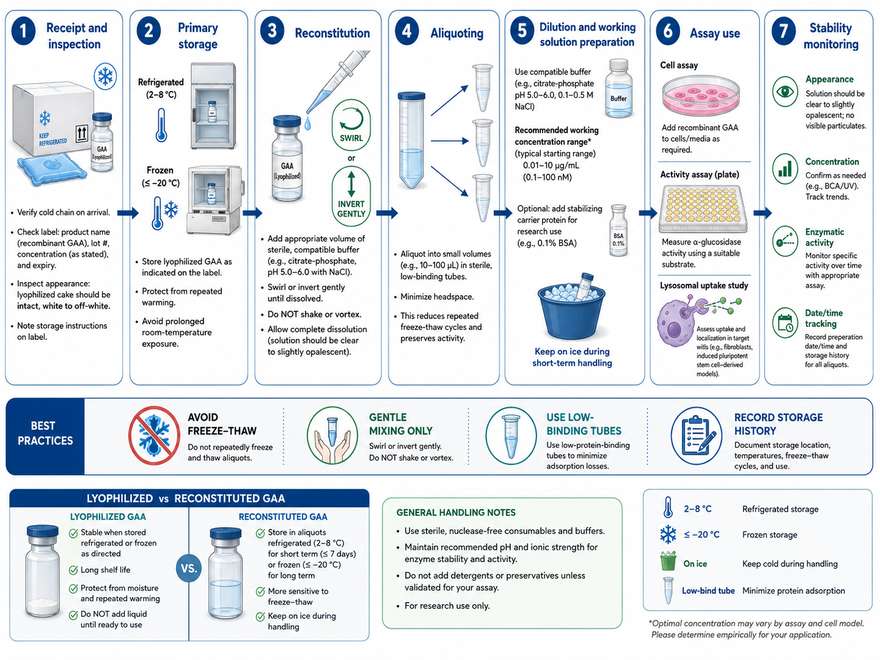
Figure 1: Recommended handling workflow for recombinant GAA, from receipt and storage to gentle reconstitution, aliquoting, dilution, assay use, and stability monitoring.
1. Why Handling Matters for Recombinant Enzymes
Recombinant enzymes are sensitive to environmental stress because catalytic function depends on a precisely folded three-dimensional structure. For GAA, handling must also preserve glycosylation-dependent uptake features and prevent the formation of aggregates or particles that can distort cellular uptake, activity, and immunogenicity studies.
| Handling Stress | Potential Molecular Effect | Possible Experimental Impact |
|---|---|---|
| Temperature excursion | Unfolding, aggregation, oxidation, or accelerated degradation | Lower activity and increased lot-to-lot variability |
| Vigorous shaking or foaming | Air-liquid interface stress and particle formation | Loss of soluble protein and inconsistent dosing |
| Repeated freeze-thaw | Cryoconcentration, pH shift, and aggregate formation | Progressive decline in activity |
| Very low concentration | Adsorption to plastic, glass, tubing, or filters | Lower effective concentration than calculated |
| Improper buffer | Charge, solubility, or catalytic environment changes | Reduced stability or altered assay response |
2. Lyophilized vs. Reconstituted GAA
Lyophilization usually improves long-term stability by limiting molecular mobility and hydrolytic reactions. Once reconstituted, however, GAA becomes more vulnerable to temperature, oxidation, agitation, microbial contamination, and surface adsorption. The usable period after reconstitution should therefore be defined by the supplier's instructions or an internally validated hold-time study.
| Form | Main Advantage | Main Risk | Recommended Practice |
|---|---|---|---|
| Lyophilized powder | Greater long-term stability | Moisture uptake and improper reconstitution | Keep container closed, dry, and within the labeled temperature range |
| Reconstituted stock | Convenient for immediate use | Aggregation, oxidation, contamination, and activity loss | Use promptly or aliquot under validated conditions |
| Diluted working solution | Ready for assay addition | Surface adsorption and dilution-dependent instability | Prepare close to use and avoid unnecessary transfers |
Instructions for a therapeutic alglucosidase alfa vial are not automatically interchangeable with those for a research-use recombinant GAA reagent. Follow the exact certificate of analysis, product insert, and formulation information supplied with the material in hand.
3. Storage Temperature Considerations
Temperature selection depends on formulation, concentration, container closure, and intended storage duration. Refrigeration is commonly used for unopened lyophilized or liquid biological products, whereas frozen storage may be appropriate for research stocks only when the supplier confirms freeze compatibility. Ultra-low temperatures do not guarantee stability if freezing creates local pH or solute concentration changes.
| Condition | Typical Use | Key Consideration |
|---|---|---|
| 2–8°C | Short- to medium-term storage of many labeled preparations | Protect from light and avoid freezing unless specifically allowed |
| −20°C | Research aliquots when validated | Use cryoprotective formulation if required; avoid frost-free cycling |
| −70°C or below | Long-term analytical reference stocks | Validate recovery after thawing and monitor concentration-dependent loss |
| Room temperature | Temporary handling or assay setup | Minimize exposure and document cumulative hold time |
4. Reconstitution Buffer Selection
The correct buffer should support solubility, preserve the native fold, and remain compatible with the planned assay. For labeled products, use only the specified diluent. For research-grade material without a fixed formulation, buffer selection should be verified by small-scale screening rather than assumed from another GAA product.
| Buffer Variable | Why It Matters | Practical Guidance |
|---|---|---|
| pH | Controls charge state, solubility, and degradation pathways | Use a range supported by supplier data and intended assay conditions |
| Ionic strength | Influences electrostatic interactions and aggregation | Avoid abrupt transfer into extremely low- or high-salt buffers |
| Surfactant | May reduce adsorption and interface-induced aggregation | Use only assay-compatible, validated concentrations |
| Stabilizing sugar or polyol | Can protect against freezing and unfolding | Confirm compatibility with activity and cell-based assays |
| Preservative | Limits microbial growth | Do not add unless validated; preservatives may affect cells or enzyme activity |
- Add diluent slowly down the vial wall.
- Allow wetting and dissolution without vigorous agitation.
- Use gentle swirling or slow inversion if permitted.
- Do not shake or create foam.
- Inspect for visible particles, discoloration, or incomplete dissolution.
5. Avoiding Freeze-Thaw Damage
Repeated freeze-thaw cycles can expose GAA to ice interfaces and locally concentrated salts, excipients, and protein. Damage is often cumulative and may not be evident by visual inspection. The preferred strategy is to prepare single-use aliquots immediately after first reconstitution or thawing.
| Best Practice | Purpose |
|---|---|
| Aliquot at a concentration appropriate for final assays | Reduces repeated dilution and transfer steps |
| Use low-binding tubes with minimal headspace | Limits adsorption and air-liquid interface exposure |
| Freeze consistently using the same protocol | Improves comparability between aliquots |
| Thaw on ice or at the validated temperature | Reduces local overheating and uncontrolled stress |
| Record the number of freeze-thaw cycles | Supports root-cause investigation when activity changes |
6. Protein Concentration and Dilution Effects
Proteins generally become more vulnerable to adsorption and interfacial loss as concentration decreases. Dilution into an assay medium can also alter pH, ionic strength, stabilizer concentration, or protein-protein interactions. This is particularly important in cell-based uptake experiments where the nominal dose may differ from the amount remaining in solution.
| Risk | Warning Sign | Mitigation |
|---|---|---|
| Surface adsorption | Low recovery at low concentration | Use low-binding consumables and minimize transfer steps |
| Precipitation after dilution | Turbidity, particles, or lower soluble protein | Perform stepwise dilution and confirm buffer compatibility |
| Stabilizer dilution | Activity decreases during assay setup | Keep excipient concentration above the validated protective threshold |
| Filter loss | Lower concentration after sterile filtration | Evaluate membrane material and protein recovery before routine use |
7. Activity Loss During Handling
Loss of GAA activity may arise from structural instability, chemical degradation, adsorption, proteolysis, or errors in assay preparation. Because catalytic assays can be influenced by pH, substrate concentration, incubation time, and normalization method, apparent activity loss should be confirmed using orthogonal measurements.
| Observed Result | Possible Cause | Follow-Up Check |
|---|---|---|
| Lower activity but unchanged protein concentration | Partial unfolding, oxidation, or catalytic-site damage | Compare structural and activity assays |
| Lower activity and lower concentration | Adsorption, precipitation, or transfer loss | Measure soluble protein and inspect containers |
| Normal cell-free activity but poor cell uptake | Loss of receptor-relevant glycans or altered aggregation state | Run CI-MPR-dependent uptake and glycan assays |
| High variability among replicates | Inconsistent thawing, mixing, timing, or dilution | Standardize workflow and operator steps |
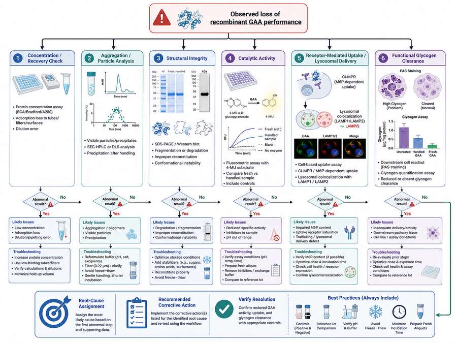
Figure 2: Integrated workflow for investigating recombinant GAA activity loss using concentration, aggregation, structural, catalytic, uptake, and glycogen-clearance measurements.
8. Stability Testing Methods
A fit-for-purpose stability program should include both physical and functional assays. No single method can distinguish every form of degradation. Long-term, accelerated, freeze-thaw, agitation, light-exposure, and in-use hold studies can be combined according to the intended research application.
| Method | Attribute Measured | Use in GAA Stability Studies |
|---|---|---|
| SEC-HPLC | Monomer, fragments, and soluble aggregates | Tracks aggregation and clipping |
| DLS or particle analysis | Hydrodynamic size and larger particles | Detects early aggregation or subvisible particles |
| SDS-PAGE / Western blot | Apparent molecular weight and integrity | Identifies fragmentation and major degradation |
| Protein concentration assay | Recoverable protein | Detects adsorption or precipitation loss |
| 4-MU enzymatic assay | Catalytic activity | Measures functional enzyme recovery |
| Cellular uptake assay | CI-MPR-dependent internalization | Confirms retained receptor-mediated delivery |
| Glycogen clearance assay | Integrated cellular function | Verifies biological correction after handling stress |
9. Practical Laboratory Checklist
| Stage | Checklist Item |
|---|---|
| Receipt | Confirm package condition, temperature indicator, lot, expiry, and documentation |
| Storage | Place at the specified temperature immediately and protect from light if required |
| Reconstitution | Use the specified diluent, add slowly, avoid shaking, and document time |
| Aliquoting | Use sterile low-binding tubes, single-use volumes, and clear labels |
| Thawing | Use a standardized thaw method and mix gently |
| Dilution | Prepare close to use, minimize transfers, and verify compatibility |
| Assay | Include a reference control and normalize timing across samples |
| Documentation | Record cumulative room-temperature time and freeze-thaw history |
10. Common Mistakes and Troubleshooting
| Common Mistake | Likely Consequence | Corrective Action |
|---|---|---|
| Shaking the vial to speed dissolution | Foaming and interface-induced aggregation | Reconstitute slowly and swirl gently |
| Using an unvalidated buffer | Precipitation or reduced activity | Return to the specified formulation or perform a buffer screen |
| Refreezing a partially used aliquot | Cumulative freeze-thaw damage | Prepare smaller single-use aliquots |
| Leaving dilute GAA in standard tubes | Adsorptive loss | Use low-binding materials and shorter hold times |
| Relying only on visual appearance | Undetected soluble aggregates or activity loss | Use analytical and functional stability-indicating assays |
| Comparing samples handled on different schedules | Artificial variability | Synchronize thaw, dilution, incubation, and readout timing |
When performance changes, compare the suspect sample with a freshly prepared reference using at least three dimensions: recoverable protein concentration, physical integrity, and functional activity. For cell-based studies, add receptor-mediated uptake or glycogen-clearance testing.
References
1. U.S. Food and Drug Administration. LUMIZYME (alglucosidase alfa) Prescribing Information, 2024.
2. European Medicines Agency. Myozyme: EPAR Product Information.
3. International Council for Harmonisation. ICH Q5C: Stability Testing of Biotechnological/Biological Products.
4. Wang, W. (2005). Protein aggregation and its inhibition in biopharmaceutics. Int J Pharm, 289(1–2): 1–30.
5. Wang, W., Nema, S., & Teagarden, D. (2010). Protein aggregation—pathways and influencing factors. Int J Pharm, 390(2): 89–99.
6. Carpenter, J. F., et al. (1997). Rational design of stable lyophilized protein formulations. Pharm Res, 14(8): 969–975.
7. Mahler, H. C., et al. (2009). Protein aggregation: pathways, induction factors and analysis. J Pharm Sci, 98(9): 2909–2934.
8. Randolph, T. W., et al. (2015). Do not drop: mechanical shock in vials causes cavitation, protein aggregation, and particle formation. J Pharm Sci, 104(2): 602–611.
9. Kamerzell, T. J., et al. (2011). Protein-excipient interactions: mechanisms and biophysical characterization applied to protein formulation development. Adv Drug Deliv Rev, 63(13): 1118–1159.
10. Manning, M. C., et al. (2010). Stability of protein pharmaceuticals: an update. Pharm Res, 27(4): 544–575.