Application of Imiglucerase in Lysosomal Storage Disease Research
Enzyme Replacement Therapy Studies and Drug Discovery Applications
Abstract
Imiglucerase, a recombinant form of human glucocerebrosidase (GCase), has become an indispensable tool in lysosomal storage disease research. This application note provides a comprehensive overview of Gaucher disease pathophysiology, the critical role of glucocerebrosidase in cellular lipid metabolism, and practical guidance for incorporating Imiglucerase products into experimental workflows for enzyme replacement therapy studies and drug discovery programs.
imiglucerase research application, lysosomal enzyme study, Gaucher disease, glucocerebrosidase, enzyme replacement therapy, sphingolipid metabolism
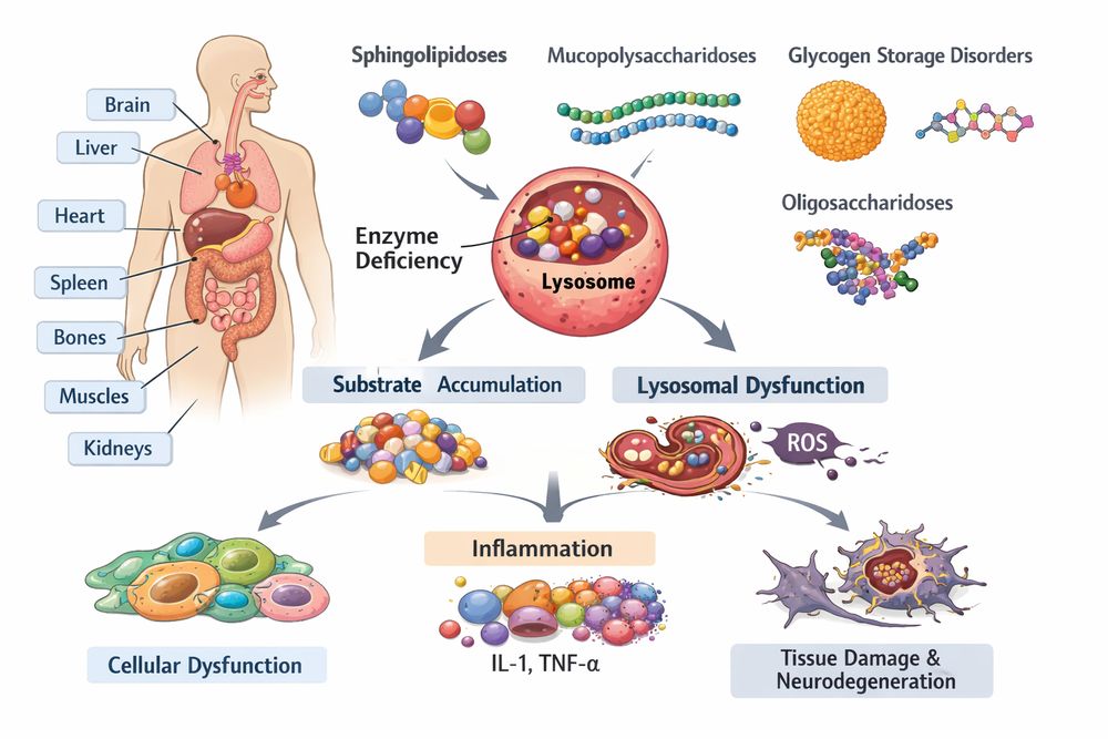
Fig1. Overview of lysosomal storage disease classification and pathophysiology
1. Overview of Lysosomal Storage Diseases
Lysosomal storage diseases (LSDs) comprise a heterogeneous group of over 70 inherited metabolic disorders characterized by defective lysosomal function. These conditions result from mutations in genes encoding lysosomal enzymes, membrane proteins, or enzyme transporters, leading to the accumulation of undegraded substrates within lysosomes.
1.1 Classification and Pathophysiology
LSDs are traditionally classified according to the nature of the accumulated substrate:
| Disease Category | Representative Disorders | Primary Enzyme Deficiency |
|---|---|---|
| Sphingolipidoses | Gaucher, Niemann-Pick, Fabry | Glucocerebrosidase, sphingomyelinase, α-galactosidase |
| Mucopolysaccharidoses | Hurler, Hunter, Sanfilippo | Iduronidase, iduronate sulfatase, heparan N-sulfatase |
| Glycogen storage | Pompe disease | Acid α-glucosidase |
| Mucolipidoses | I-cell disease | N-acetylglucosamine phosphotransferase |
The pathological cascade in LSDs extends beyond simple substrate accumulation. Secondary mechanisms include inflammatory responses through macrophage and microglia activation, autophagic dysfunction via impaired autophagosome-lysosome fusion, calcium dysregulation from lysosomal membrane permeabilization, and mitochondrial dysfunction causing oxidative stress and energy failure.
1.2 Gaucher Disease: A Prototype LSD
Gaucher disease represents the most common LSD, with an incidence of approximately 1 in 50,000 to 1 in 100,000 in the general population, and higher prevalence in Ashkenazi Jewish populations (1 in 850). The disease results from biallelic mutations in the GBA1 gene encoding lysosomal glucocerebrosidase.
Three clinical subtypes are recognized:
- Type 1 (Non-neuronopathic): Visceral manifestations without central nervous system involvement
- Type 2 (Acute neuronopathic): Rapid neurodegeneration with early mortality
- Type 3 (Chronic neuronopathic): Progressive neurodegeneration with slower course
2. Role of Glucocerebrosidase in Lipid Metabolism
2.1 Enzymatic Function and Substrate Specificity
Glucocerebrosidase (acid β-glucosidase, EC 3.2.1.45) is a lysosomal hydrolase responsible for the cleavage of glucose from glucosylceramide (GlcCer), a critical step in glycosphingolipid catabolism. The enzyme requires the sphingolipid activator protein saposin C for optimal activity in vivo.
Glucosylceramide + H₂O → Ceramide + Glucose (catalyzed by GCase)
2.2 Cellular Consequences of Enzyme Deficiency
In Gaucher disease, glucocerebrosidase deficiency leads to the progressive accumulation of GlcCer and its deacylated derivative glucosylsphingosine (GlcSph) primarily within macrophages. These "Gaucher cells" exhibit characteristic morphology including cytoplasmic striations (tubular inclusions of accumulated substrate), eccentric nuclei compressed by distended lysosomes, and enhanced CD68/CD163 expression indicating activated macrophage phenotype.
The metabolic block creates a ripple effect throughout cellular lipid homeostasis, including sphingolipid rheostat disruption (altered ceramide/sphingosine-1-phosphate balance), cholesterol trafficking defects (secondary NPC1-like phenotype), and membrane raft perturbation causing impaired receptor signaling.
2.3 Connection to Parkinson's Disease
Recent research has established a significant association between GBA1 mutations and Parkinson's disease (PD). Heterozygous GBA1 carriers have a 5-20 fold increased risk of developing PD, positioning glucocerebrosidase as a critical therapeutic target for neurodegenerative disorders beyond LSDs.
3. Research Models for Gaucher Disease
3.1 Cellular Models
Primary patient-derived cells provide physiologically relevant platforms for Imiglucerase research application:
| Cell Type | Source | Applications |
|---|---|---|
| Fibroblasts | Skin biopsies | Enzyme activity assays, substrate accumulation studies |
| Macrophages | PBMC differentiation | Phagocytosis, cytokine profiling |
| iPSC-derived neurons | Reprogrammed fibroblasts | Neurodegeneration mechanisms |
| Hepatocytes | Liver biopsies | Glycolipid metabolism studies |
CRISPR/Cas9 technology enables the generation of isogenic cell lines with specific GBA1 mutations (N370S, L444P, D409H), providing standardized platforms for high-throughput screening and mechanistic studies.
3.2 Animal Models
- Gba1 knockout mice: Embryonic lethal, limiting utility
- Gba1 hypomorphic mice: Viable with visceral and neurological phenotypes
- Gba1 conditional knockout: Tissue-specific deletion for mechanistic studies
- Zebrafish models: Optimal for high-throughput drug screening
3.3 Biochemical Assays
Standardized protocols for assessing glucocerebrosidase activity include fluorometric assay using 4-methylumbelliferyl-β-D-glucopyranoside substrate, mass spectrometry for quantification of GlcCer and GlcSph species, Western blotting for GCase protein levels and processing, and immunofluorescence for lysosomal colocalization studies.
4. Application of Recombinant Imiglucerase
4.1 Molecular Characteristics
Imiglucerase is a recombinant DNA-produced analog of human β-glucocerebrosidase, manufactured in Chinese hamster ovary (CHO) cells. The enzyme undergoes post-translational modification to expose terminal mannose residues, facilitating uptake via the mannose receptor on macrophages.
- Molecular weight: ~60 kDa (glycosylated)
- Specific activity: >800 U/mg protein
- Carbohydrate content: ~8% (mannose-terminated oligosaccharides)
- Storage: 2-8°C, stable for 24 months
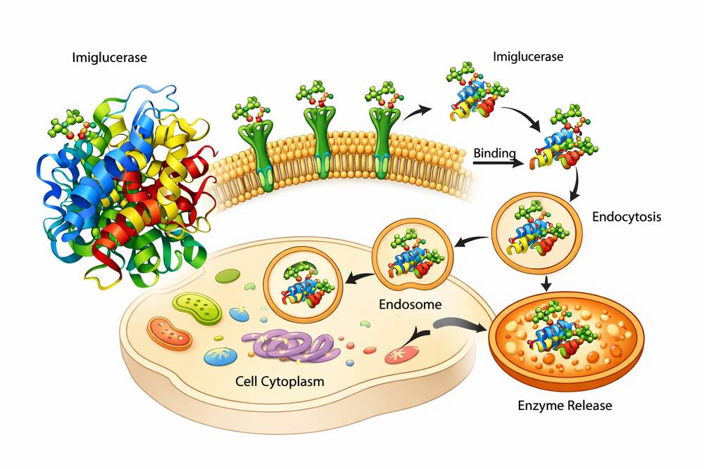
Fig2. Molecular structure of Imiglucerase and cellular uptake mechanism via mannose receptors
4.2 Research Applications
Researchers utilizing Imiglucerase products can address multiple experimental objectives:
A. Enzyme Replacement Therapy (ERT) Optimization
- Dose-response relationships in cellular models
- Uptake kinetics in different cell types
- Biodistribution studies using labeled enzyme
B. Comparative Studies
- Efficacy comparison with next-generation ERT (velaglucerase alfa, taliglucerase alfa)
- Substrate reduction therapy combinations
- Chaperone-mediated enzyme enhancement
C. Mechanistic Investigations
- Lysosomal enzyme trafficking pathways
- Immune responses to exogenous enzyme
- Biomarker modulation (chitotriosidase, CCL18)
4.3 Experimental Considerations
When designing experiments with lysosomal enzyme study concentrations of Imiglucerase, consider the following parameters:
| Parameter | Optimization Range | Notes |
|---|---|---|
| Concentration | 1-100 U/mL | Cell-type dependent uptake |
| Treatment duration | 24-72 hours | Substrate clearance kinetics |
| Temperature | 37°C | Optimal enzyme activity |
| pH | 5.0-6.0 | Lysosomal targeting verification |
5. Experimental Workflow
5.1 Cell-Based Enzyme Uptake Assay
This protocol demonstrates the application of Imiglucerase in quantifying cellular enzyme internalization and activity restoration.
Materials Required:
- Gaucher patient fibroblasts or macrophages
- Imiglucerase (reconstituted in sterile PBS)
- Fluorogenic substrate (4-MU-β-D-glucopyranoside)
- Sodium taurocholate (activator)
- McIlvaine buffer (pH 5.4)
Procedure:
- Cell Preparation: Seed cells at 5×10⁴ cells/well in 24-well plates; culture for 24 hours in complete medium
- Enzyme Treatment: Replace medium with serum-free DMEM containing Imiglucerase (0, 1, 5, 10, 50 U/mL); incubate for 4-24 hours at 37°C, 5% CO₂
- Cell Harvesting: Wash cells twice with ice-cold PBS; lyse in 200 μL distilled water by freeze-thaw cycling
- Enzyme Activity Assay: Mix 50 μL cell lysate with 100 μL substrate solution; incubate at 37°C for 60 minutes; terminate reaction with 1 mL 0.2 M glycine buffer (pH 10.7); measure fluorescence (Ex/Em: 365/450 nm)
- Data Analysis: Normalize to total protein content; calculate specific activity (nmol/h/mg protein); determine dose-response curves and EC₅₀ values
5.2 Substrate Clearance Quantification
Lipid Extraction and Analysis:
- Treat cells with lysosomal enzyme study concentrations of Imiglucerase for 48-72 hours
- Harvest cells and extract lipids using chloroform:methanol (2:1)
- Analyze GlcCer species by LC-MS/MS
- Normalize to phospholipid phosphate or protein content
6. Expected Results
6.1 Enzyme Activity Restoration
Treatment of Gaucher fibroblasts with Imiglucerase typically yields:
- Dose-dependent activity increase: 5-10 fold restoration at 10 U/mL
- Time-dependent accumulation: Maximal activity at 24-48 hours
- Cell-type variability: Macrophages show 3-5 fold higher uptake than fibroblasts
| Imiglucerase (U/mL) | Enzyme Activity (% of Normal) | GlcCer Reduction (%) |
|---|---|---|
| 0 | 5-15 | 0 |
| 1 | 25-35 | 15-25 |
| 5 | 55-70 | 40-55 |
| 10 | 80-95 | 65-80 |
| 50 | 100-120 | 85-95 |
6.2 Morphological Improvements
Microscopic evaluation should demonstrate reduced lysosomal swelling (electron microscopy verification), decreased CD68 staining (immunohistochemical quantification), and normalized cytokine profiles (IL-1β, IL-6, TNF-α reduction).
6.3 Biomarker Modulation
In macrophage models, expect chitotriosidase activity reduction of 50-70% after 72 hours, significant decrease in secreted CCL18/PARC chemokine, and ferritin normalization indicating iron homeostasis restoration.
The integration of high-quality Imiglucerase products into lysosomal storage disease research workflows enables precise investigation of enzyme replacement mechanisms, substrate clearance kinetics, and therapeutic optimization. As the field advances toward combination therapies and novel delivery systems, recombinant glucocerebrosidase remains a fundamental tool for both basic research and translational applications.
References
1. Platt FM, et al. (2018) Lysosomal storage diseases. Nature Reviews Disease Primers 4:27
2. Stirnemann J, et al. (2017) The diagnosis and management of Gaucher disease. Orphanet Journal of Rare Diseases 12:72
3. Vaccaro AM, et al. (2010) Saposins and their interaction with lipids. Neurochemical Research 35:1878-1884
4. Sidransky E, et al. (2009) Multicenter analysis of glucocerebrosidase mutations in Parkinson's disease. New England Journal of Medicine 361:1651-1661
5. Grabowski GA, et al. (2014) Enzyme therapy for Gaucher disease: The first 25 years. Blood Reviews 28:357-363