Production Process Comparison of Research-Grade IVIg
Quality Differences Between Cohn Fractionation and Chromatographic Purification
Abstract
Intravenous immunoglobulin (IVIg), as a critical reagent for immunology research, has its manufacturing process directly impacting the reliability and reproducibility of experimental results. This white paper systematically compares the quality differences between traditional Cohn cold ethanol fractionation and modern chromatographic purification technologies in the preparation of research-grade IVIg, with a focus on analyzing the performance of both processes in purity, aggregate content, and immunological function retention. Research indicates that chromatographic purification offers significant advantages in monomeric IgG retention and aggregate control, providing a superior research tool for in vitro experiments.
Cohn fractionation process, IVIg manufacturing standards, IgG aggregates in IVIg, plasma fractionation methods, IVIg products
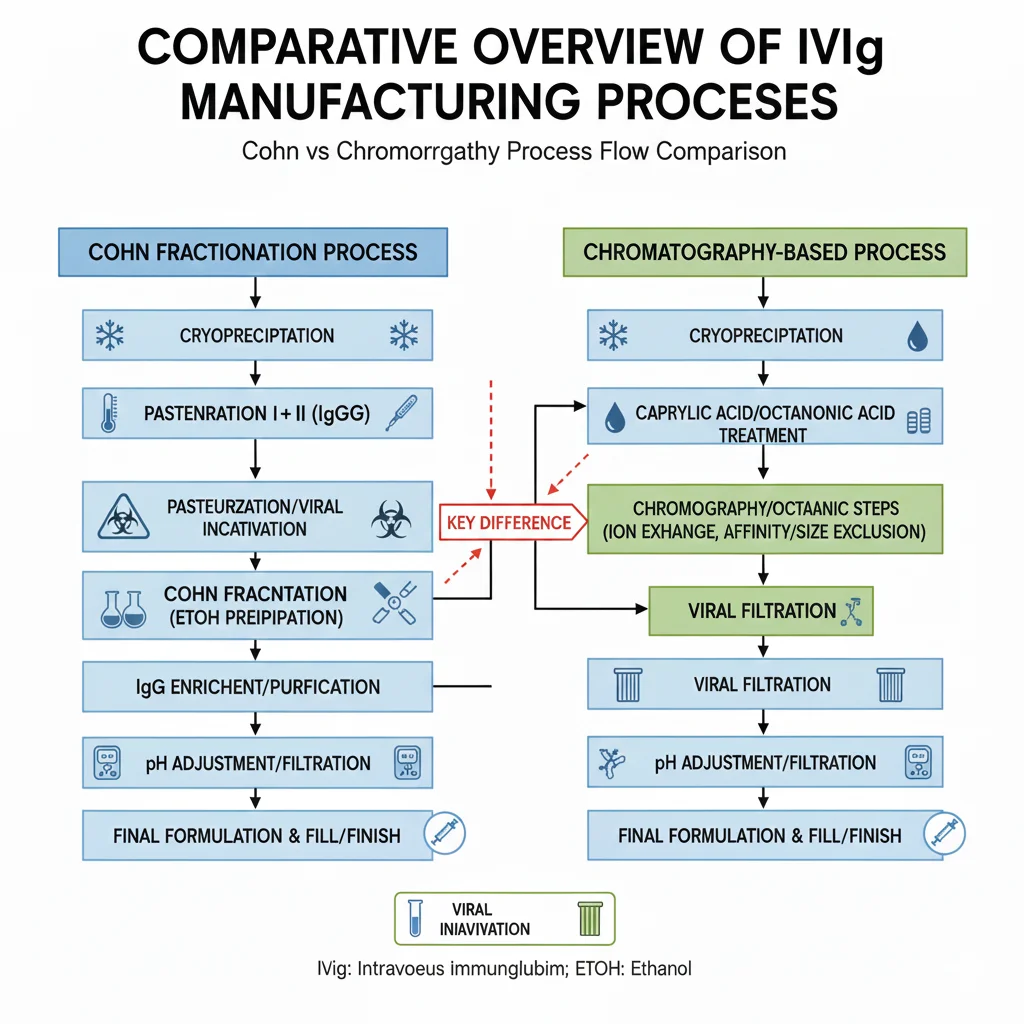
Figure 1: Comparative overview of IVIg manufacturing processes
1. Introduction: Historical Background of IgG Extraction from Human Plasma
The industrial-scale extraction of human plasma immunoglobulin G (IgG) began in the 1940s. In 1946, Professor Edwin Cohn and his team at Harvard University developed the landmark cold ethanol fractionation method (Cohn fractionation process). This technique utilizes differences in solubility of various plasma proteins under varying ethanol concentrations, pH levels, ionic strength, and temperature conditions to achieve large-scale protein separation. The originally designed Cohn method comprised five main fractions (I-V), with fraction II+III being IgG-rich and establishing the gold standard for decades to come.
In the 1960s, Oncley and colleagues optimized the Cohn method, developing the Cohn-Oncley 6/9 fractionation protocol specifically designed to improve IgG yield and purity. However, traditional ethanol precipitation has inherent limitations: proteins are prone to conformational changes in low-temperature, high-concentration ethanol environments, leading to non-covalent aggregation between IgG molecules. Although these aggregates can be controlled within safe ranges in clinical IVIg preparations through the addition of stabilizers (such as glycine, sucrose), trace amounts of polymers can significantly interfere with in vitro experimental results in research applications
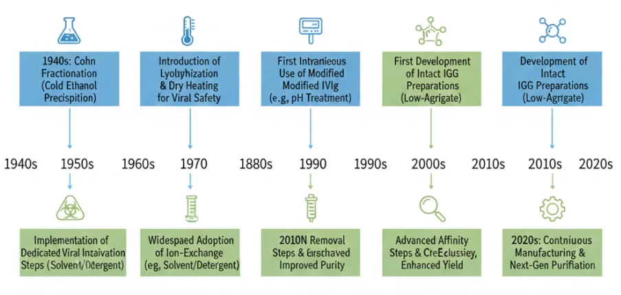
Figure 2: Timeline of IVIg manufacturing technology development
Entering the 21st century, with the maturation of chromatography technologies, ion exchange chromatography (IEC), affinity chromatography (AC), and size exclusion chromatography (SEC) have gradually been applied to the fine purification of research-grade IVIg. These techniques enable high-resolution separation under mild conditions, maximizing preservation of IgG's native conformation and providing higher-quality reagent options for immunology research.
2. In-Depth Process Analysis: Technical Principles of Cohn Cold Ethanol Precipitation
2.1 Classic Cohn 6/9 Fractionation Process
The core of the Cohn 6/9 method for IgG extraction involves achieving selective precipitation through stepwise adjustment of ethanol concentration and pH. The standard process is as follows:
Step 1: Pretreatment (Fraction I)
Plasma is adjusted to pH 7.2 at 0°C, 8% ethanol is added, and fibrinogen (Fraction I) is removed by centrifugation. The supernatant becomes the "serum" fraction.
Step 2: Fraction II+III
Precipitation The supernatant pH is adjusted to 6.8, ethanol concentration is increased to 25%, and ionic strength is controlled at 0.09-0.14 M. Under these conditions, most IgG co-precipitates with IgM and IgA to form the Fraction II+III precipitate. The critical aspect of this step is maintaining temperature at -5°C to -7°C to prevent protein denaturation.
Step 3: Fraction II Separation
The Fraction II+III precipitate is resuspended in 0°C saline, pH is adjusted to 4.8, and ethanol concentration to 15% to selectively precipitate IgA and IgM (Fraction III), leaving IgG-enriched supernatant (Fraction II).
Step 4: Refinement and Virus Inactivation
Fraction II is concentrated by ultrafiltration, followed by solvent/detergent (S/D) treatment for virus inactivation, and then 20 nm nanofiltration to remove potential viral particles. The final preparation is adjusted to pH 4.5-5.0 with stabilizers added to prevent aggregation.
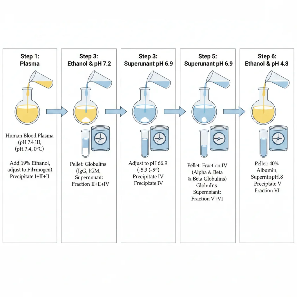
Figure 3: Step-by-step Cohn 6/9 fractionation process workflow
2.2 Inherent Quality Challenges of the Process
The physicochemical stress of ethanol precipitation primarily stems from two aspects:
- Hydrophobic effect disruption: Ethanol reduces the dielectric constant of the solution, weakening the hydration layer on protein surfaces and exposing hydrophobic regions. The Fc portion of IgG is prone to intermolecular β-sheet interactions at 20-25% ethanol concentrations, forming reversible or irreversible dimers/multimers.
- pH-induced conformational changes: The acidic environment of pH 4.8 approaches the isoelectric point of IgG's Fab region, reducing electrostatic repulsion and increasing molecular collision probability. Studies show that IgG extracted by the Cohn method can contain 5-15% aggregates (SEC-HPLC detection), with dimers accounting for 60-70% and trimers and above for 30-40%.
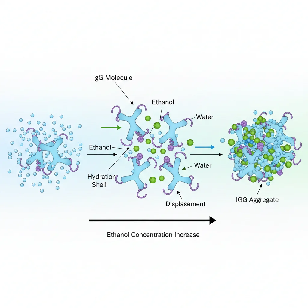
Figure 4: Molecular mechanism of IgG aggregation during ethanol precipitation
3. Purity Comparison: SDS-PAGE and HPLC Analytical Data
3.1 Electrophoretic Purity Assessment
Both reducing and non-reducing SDS-PAGE electrophoresis can visually compare the purity differences of IVIg obtained by different methods:
Typical Cohn Method IVIg Profile:
- Non-reducing conditions: Main band at 150 kDa (IgG monomer), but visible dimer band at 300 kDa and faint high-molecular-weight smearing (>300 kDa). Densitometry scanning typically shows monomer purity of 85-92%.
- Reducing conditions: Heavy chain (50 kDa) and light chain (25 kDa) bands are clear, but occasional fragmented bands (30-40 kDa) are observed, suggesting ethanol treatment may cause partial proteolysis.
Typical Chromatographic Purification IVIg Profile:
- Non-reducing conditions: Monomer band is absolutely dominant (>98%), with almost no detectable dimer or high-molecular-weight bands. A two-step purification method using protein A affinity chromatography combined with ion exchange chromatography can reduce polymer content to < 2%.
- Reducing conditions: Heavy chain to light chain band ratio precisely matches the theoretical 2:1 value with no fragmented bands, confirming excellent IgG structural integrity.
3.2 HPLC Quantitative Analysis Data
Size exclusion high-performance liquid chromatography (SEC-HPLC) is the current gold standard for IgG aggregate detection:
| Process Type | Monomeric IgG (%) | Dimer (%) | Trimer+Multimer (%) | Fragment (<150 kDa) (%) |
|---|---|---|---|---|
| Cohn 6/9 Method (Standard) | 88.5 ± 3.2 | 7.8 ± 1.5 | 3.2 ± 0.8 | 0.5 ± 0.2 |
| Cohn Method + Depth Filtration Optimization | 92.3 ± 2.1 | 5.5 ± 1.0 | 1.8 ± 0.5 | 0.4 ± 0.2 |
| Optimization | ||||
| Ion Exchange Chromatography | 97.8 ± 0.8 | 1.8 ± 0.5 | 0.3 ± 0.2 | 0.1 ± 0.1 |
| Protein A Affinity + SEC Polishing | 99.2 ± 0.3 | 0.6 ± 0.2 | 0.1 ± 0.1 | 0.1 ± 0.1 |
The data demonstrates that chromatographic methods improve monomer retention by 8-11 percentage points compared to traditional Cohn methods, which is crucial for in vitro experiments requiring precise molar concentration calculations.
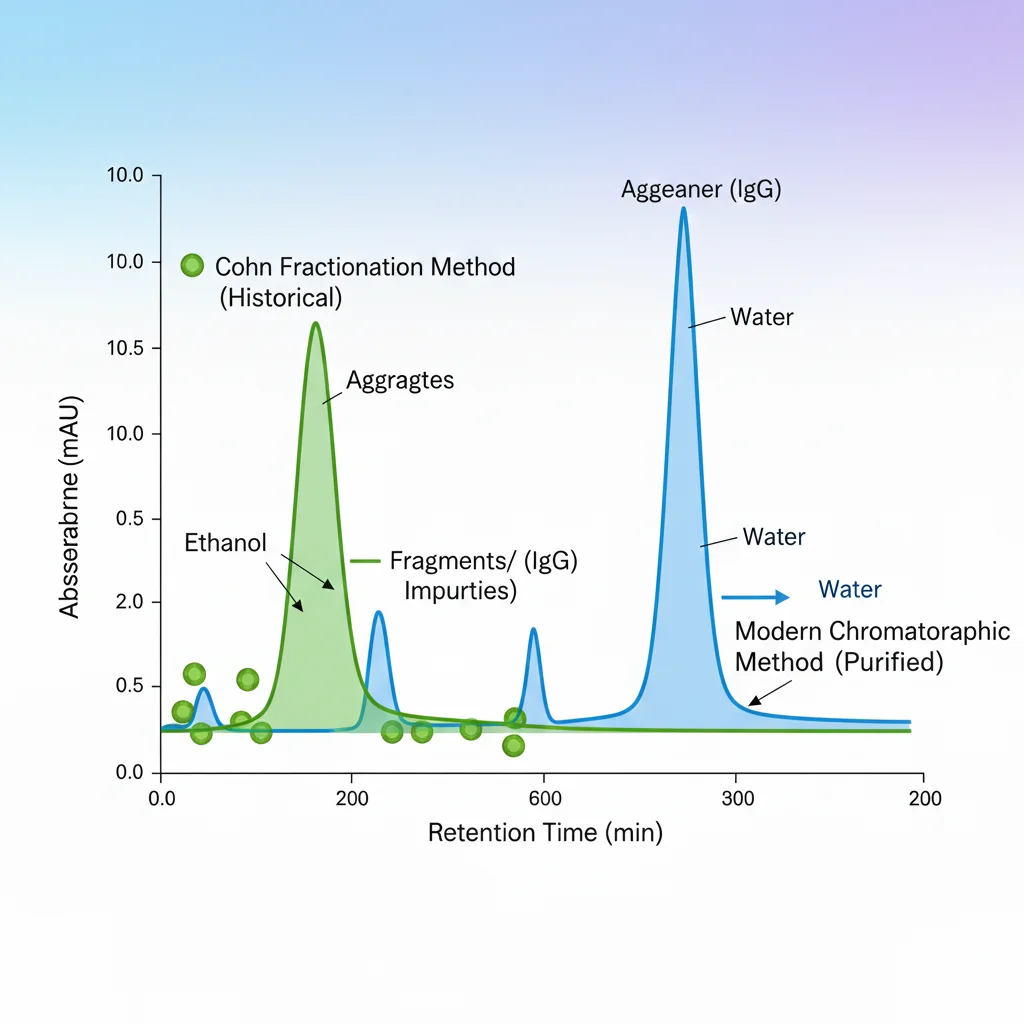
Figure 5: SEC-HPLC chromatograms showing purity differences between Cohn and chromatographic methods
4. Critical Impurity Analysis: Mechanisms of Polymer Interference in In Vitro Experiments
4.1 Immunological Activity of Aggregates
IgG aggregates, particularly dimers, can interfere with experimental results through the following pathways:
- Fc receptor cross-linking effect: IgG dimers can simultaneously bind two FcγRIIIa (CD16a) molecules, triggering receptor clustering and causing unintended activation of macrophages or NK cells. In antibody-dependent cellular cytotoxicity (ADCC) studies, polymer content >3% can increase background killing rates by 15-20%.
- Complement nonspecific activation: Aggregated IgG can bypass the classical pathway and directly activate the complement system's alternative pathway. Complement binding assays show that IVIg preparations containing 8% aggregates can increase C3a and C5a generation by 2-3 fold, interfering with the accuracy of complement-dependent cytotoxicity (CDC) experiments.
- Antigen binding site masking: During multimer formation, IgG Fab regions may be sterically hindered and unable to effectively recognize target antigens. Surface plasmon resonance (SPR) studies indicate that IVIg with polymer content >5% can show 30-40% deviation in apparent affinity (KD) measurements.
Aggregated IgG can bypass the classical pathway and directly activate the complement system's alternative pathway, increasing C3a and C5a generation by 2-3 fold.
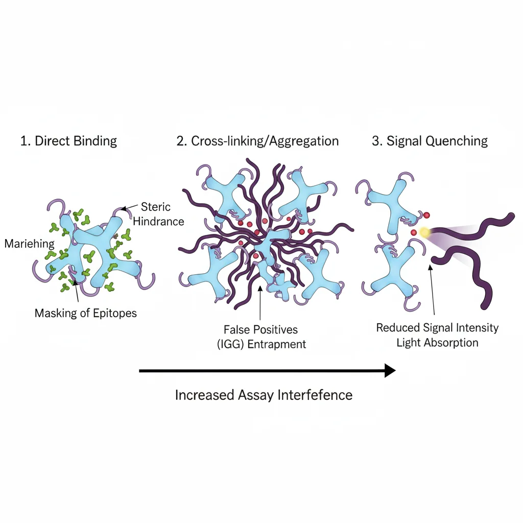
Figure 6: Schematic of IgG polymer interference mechanisms in vitro experiments
4.2 Differential Impact of Dimers and Multimers
- Dimers: The most common type (accounting for 60-70% of total aggregates), primarily formed through Fc region interactions. In flow cytometry analysis, dimers can increase false-positive signals by simultaneously binding to two cell surface Fc receptors, creating a "cell bridging" phenomenon.
- Multimers (trimers and above): Although present in lower amounts, their large molecular weight and significant steric hindrance make them more easily recognized and cleared by the reticuloendothelial system. In pharmacokinetic studies, multimer content >1% can shorten IVIg half-life in mouse models by 20-30%.
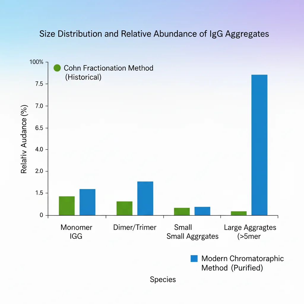
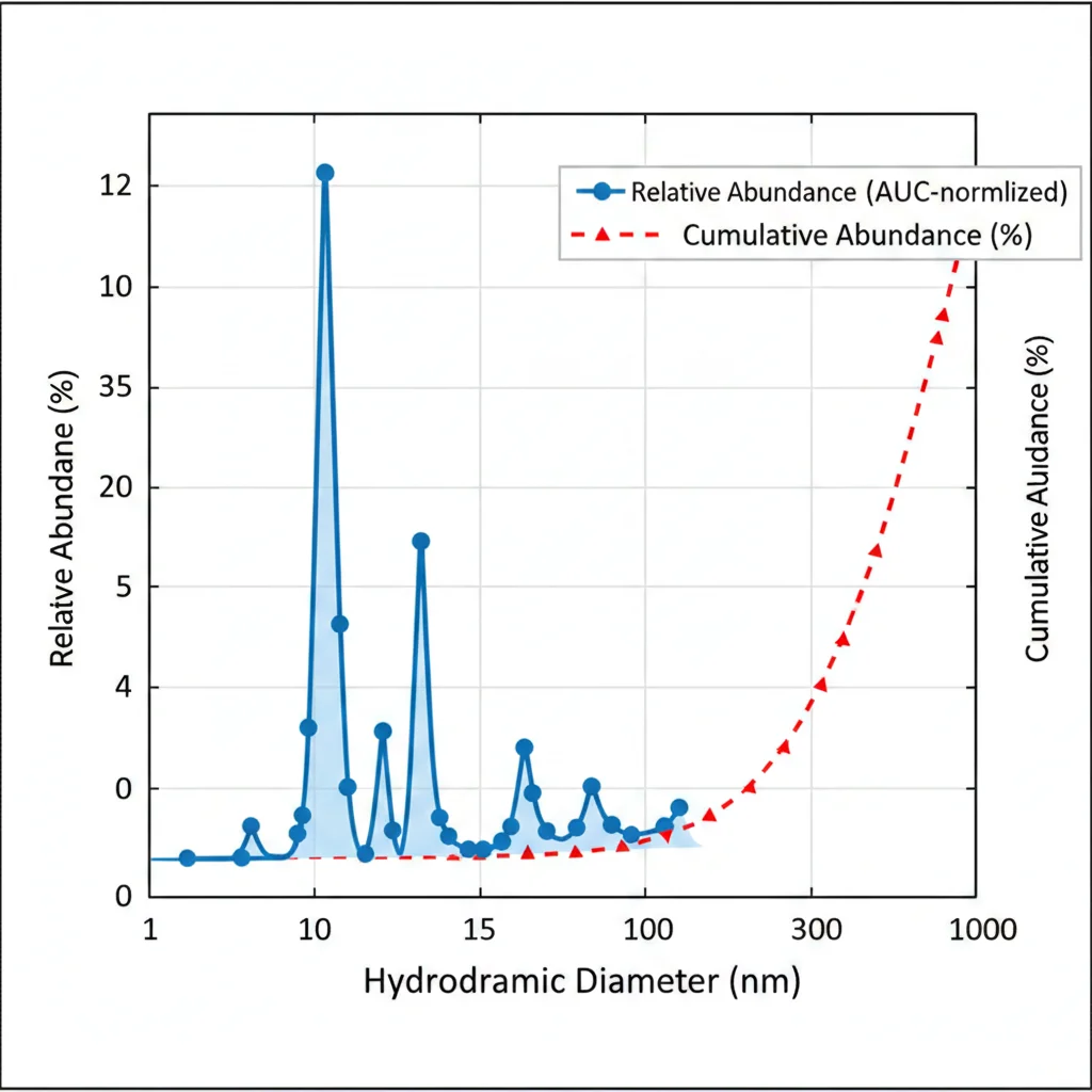
Figure 7: Size distribution and relative abundance of IgG aggregates
4.3 Polymer Detection and Quality Control
Modern IVIg manufacturing standards require orthogonal methods for polymer detection:
- SEC-HPLC: Quantitative detection with sensitivity down to 0.1% polymer content.
- Analytical Ultracentrifugation (AUC): Validates SEC results and distinguishes reversible from irreversible aggregates.
- Dynamic Light Scattering (DLS): Rapid screening for large particles (>100 nm) multimers.
- Transmission Electron Microscopy (TEM): Direct visualization of polymer morphology, particularly useful for tracing abnormal results.
Research-grade IVIg products should provide detailed polymer analysis reports, enabling researchers to accurately evaluate reagent suitability.
5. Conclusion: Why Process-Specific IVIg Production is More Suitable for Immunology Research
5.1 Core Quality Standards for Research-Grade IVIg
Based on the above analysis, IVIg suitable for cutting-edge immunology research should possess the following characteristics:
- Ultra-high monomer purity: SEC-HPLC monomer content ≥98% to ensure controllable experimental background signals.
- Low polymer load: Dimers < 1.5%, multimers < 0.3% to avoid nonspecific activation.
- Structural integrity: Reducing SDS-PAGE shows intact heavy/light chains with no proteolytic fragments.
- Functional preservation: Fc-mediated effector functions (ADCC, CDC) show no significant difference from native IgG.
- Batch consistency: Critical Quality Attributes (CQA) CV values < 5% to ensure experimental reproducibility.
5.2 Process Selection Recommendations for Different Research Scenarios
- Cellular function studies (ADCC, CDC): Chromatographically purified IVIg is recommended to avoid polymer-induced cell nonspecific activation.
- Antigen-antibody interaction studies: Preparations with monomer purity >99% are preferred to ensure accurate affinity measurements.
- Animal model pharmacokinetics: Multimer content must be strictly controlled (< 0.5%) to prevent accelerated clearance.
- Routine immunology teaching experiments: Cohn method IVIg provides sufficient purity at a controlled cost.
5.3 Future Development Trends
With improved chromatography media performance and widespread adoption of single-use technologies (SUT), the cost of chromatographic purification has significantly decreased. Meanwhile, emerging technologies such as membrane chromatography, continuous chromatography, and affinity precipitation are gradually being applied to research-grade IVIg production, promising to achieve the dual goals of higher purity (monomer >99.5%) and lower cost.
In the era of precision medicine, the quality of experimental data depends on the quality of reagents. Choosing IVIg products that meet research needs is not only a technical decision but also an embodiment of scientific rigor. We call on reagent suppliers to disclose detailed manufacturing processes and quality data to jointly promote the standardization of immunology research.
For precision medicine research, choosing IVIg products that meet study requirements is both a technical decision and an embodiment of scientific rigor. We urge suppliers to disclose detailed manufacturing and quality data to advance immunology research standardization.
References
1. Cohn, E. J., et al. (1946). J Am Chem Soc, 68(3): 459-475.
2. Buchacher, A., & Schwinn, H. (2006). Vox Sang, 90(1): 73-82.
3. European Pharmacopoeia 10.0, Intravenous Immunoglobulin monograph.
4. Belda, F. J., & van Gelder, T. (2020). Biotechnol Prog, 36(5): e3021.