
Thyroid-Stimulating Hormone Biology: Regulation, Signaling, and Thyroid Function
From Pituitary Secretion to TSH Receptor Activation and Thyroid Hormone Biosynthesis
Abstract
Thyroid-stimulating hormone (TSH), also called thyrotropin, is the principal pituitary signal that coordinates thyroid growth, differentiated thyroid function, and thyroid hormone production. Its secretion is controlled by hypothalamic thyrotropin-releasing hormone (TRH), circulating thyroid hormones, circadian timing, nutritional state, illness, and multiple central and peripheral modulators. At the thyroid gland, TSH binds the TSH receptor (TSHR) on follicular cells and activates signaling networks that support iodide uptake, thyroglobulin synthesis, iodination, hormone release, and cell survival.
This white paper reviews the biological organization of the hypothalamic-pituitary-thyroid (HPT) axis, the molecular events downstream of TSHR activation, and the experimental systems commonly used to study TSH responses. Particular attention is given to the differences between physiological TSH exposure and laboratory stimulation, because hormone source, glycosylation, species compatibility, dose, exposure pattern, receptor density, and assay timing can substantially influence observed activity.
thyroid-stimulating hormone biology, TSH regulation, TSH receptor signaling, hypothalamic-pituitary-thyroid axis, TRH-mediated TSH secretion, iodide uptake, thyroglobulin synthesis, T3 and T4 feedback, thyroid follicular cells, bovine thyroid-stimulating hormone (bTSH)
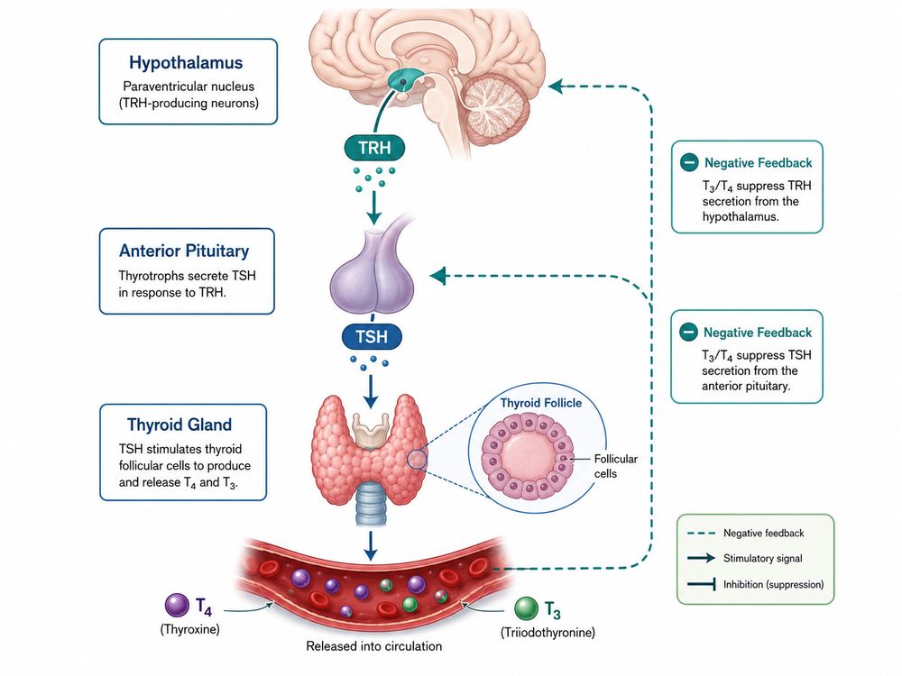
Fig 1. Integrated view of the hypothalamic-pituitary-thyroid axis and T3/T4-mediated negative feedback.
1. What Is Thyroid-Stimulating Hormone?
TSH is a heterodimeric glycoprotein hormone produced primarily by thyrotroph cells in the anterior pituitary. It consists of a common alpha subunit shared with luteinizing hormone, follicle-stimulating hormone, and human chorionic gonadotropin, plus a hormone-specific beta subunit that provides most of the receptor selectivity. The two subunits must assemble correctly and undergo appropriate glycosylation before biologically active TSH is secreted.
TSH should not be interpreted as a simple on/off switch. Its biological activity reflects concentration, pulse frequency, glycan composition, receptor abundance, ligand-receptor residence time, and the intracellular state of the target cell. Glycosylation can influence receptor activation, metabolic clearance, immunoreactivity, and the relationship between measured TSH concentration and functional potency. Consequently, equal masses or immunoreactive concentrations of two TSH preparations may not produce identical responses in a cell-based assay.
In experimental work, purified pituitary-derived or recombinant TSH can be used to stimulate TSHR-dependent pathways. A TSH research reagent may be selected for receptor activation, cAMP measurement, iodide uptake studies, thyroid differentiation assays, or thyroid hormone synthesis models. The dose should be defined by an assay-specific dose-response curve rather than transferred uncritically from another cell type or species.
2. TSH Production in the Anterior Pituitary
Anterior pituitary thyrotrophs synthesize the common glycoprotein alpha subunit and the TSH beta subunit, assemble them in the secretory pathway, and release mature TSH into the circulation. TSH beta expression is especially important for hormone specificity and is tightly regulated by stimulatory TRH signaling and inhibitory thyroid hormone feedback. Thyrotroph differentiation and gene expression also depend on transcriptional regulators that establish pituitary lineage and endocrine responsiveness.
TSH secretion occurs in pulses superimposed on a circadian rhythm. In humans, secretion generally rises during the evening and reaches its highest levels during the night, although sleep itself modifies the amplitude of the nocturnal rise. The resulting TSH profile is therefore shaped by both the central circadian pacemaker and sleep-wake state.
Pituitary output can change without a primary alteration in the thyroid gland. Energy restriction, severe systemic illness, medications, glucocorticoid signaling, somatostatin, dopamine, and changes in central deiodinase activity can alter TRH or TSH production. Experimental interpretation should therefore distinguish thyroid-intrinsic responsiveness from upstream changes in hypothalamic or pituitary regulation.
3. The Hypothalamic-Pituitary-Thyroid Axis
The HPT axis is a hierarchical feedback system that maintains thyroid hormone availability within a regulated physiological range. Hypophysiotropic TRH neurons in the paraventricular nucleus of the hypothalamus release TRH into the median eminence. TRH then travels through the hypothalamic-hypophyseal portal circulation to the anterior pituitary, where it stimulates thyrotrophs. Pituitary TSH enters the systemic circulation and activates TSHR on thyroid follicular cells. The thyroid produces predominantly thyroxine (T4), along with smaller amounts of triiodothyronine (T3), and peripheral tissues convert T4 to active T3 or inactive metabolites according to local requirements.
| Axis Level | Primary Signal | Main Target | Representative Biological Output |
|---|---|---|---|
| Hypothalamus | TRH | Anterior pituitary thyrotrophs | TSH synthesis, maturation, and secretion |
| Anterior pituitary | TSH | Thyroid follicular cells | TSHR signaling, thyroid differentiation, and hormone biosynthesis |
| Thyroid gland | T4 and T3 | Pituitary, hypothalamus, and peripheral tissues | Metabolic regulation and negative feedback |
| Peripheral tissues | Deiodinase-controlled T3 availability | Local thyroid hormone receptors | Tissue-specific thyroid hormone action |
A change in serum TSH can result from altered hypothalamic drive, pituitary sensitivity, thyroid hormone feedback, thyroid gland function, assay interference, or combinations of these factors. TSH is therefore most informative when interpreted together with T4/T3 measurements and the experimental context.
4. TRH-Mediated Regulation of TSH Secretion
TRH binds the TRH receptor on pituitary thyrotrophs and primarily activates a Gq/11-phospholipase C pathway. This produces inositol trisphosphate and diacylglycerol, increases intracellular calcium, activates protein kinase C, and promotes exocytosis of stored TSH. TRH also supports longer-term TSH subunit gene expression and can influence post-translational maturation of the secreted hormone.
The magnitude of the TSH response depends on prior thyroid hormone exposure, receptor availability, secretory reserve, pulse timing, and the physiological state of the pituitary. A brief TRH pulse can produce an acute secretory response, whereas prolonged or repeated stimulation can change synthesis, glycosylation, receptor responsiveness, and intracellular signaling. This distinction is relevant when comparing rapid secretion assays with longer gene-expression experiments.
TRH neurons integrate inputs related to temperature, nutritional status, stress, inflammation, and energy balance. During fasting or severe illness, central mechanisms can reduce TRH drive even when peripheral thyroid hormone concentrations are low or changing. Such adaptive responses illustrate why the HPT axis is not governed by thyroid hormone feedback alone.
5. TSH Binding to Thyroid Follicular Cells
TSHR is a cell-surface G protein-coupled receptor characterized by a large extracellular leucine-rich repeat domain that binds TSH and a seven-transmembrane domain that transduces the signal. In differentiated thyroid tissue, TSHR is expressed on the basolateral membrane of polarized follicular cells, positioning the receptor to detect circulating TSH while the apical surface faces the colloid-filled follicular lumen.
TSH receptor activation most commonly engages Gs-mediated activation of adenylyl cyclase, accumulation of cyclic AMP (cAMP), and activation of protein kinase A. This pathway regulates genes required for differentiated thyroid function, including components involved in iodide transport and thyroglobulin production. Depending on receptor density, ligand concentration, cell background, and experimental conditions, TSHR can also engage Gq/11-phospholipase C signaling, intracellular calcium responses, kinase cascades, receptor internalization, and beta-arrestin-associated processes.
TSH-dependent proliferation is highly context dependent. In many cultured thyrocyte systems, cAMP signaling cooperates with insulin or insulin-like growth factor signaling rather than acting as a fully independent mitogenic input. A growth response should therefore not be assumed to represent the same signaling state as a differentiated response such as NIS induction or iodide uptake.
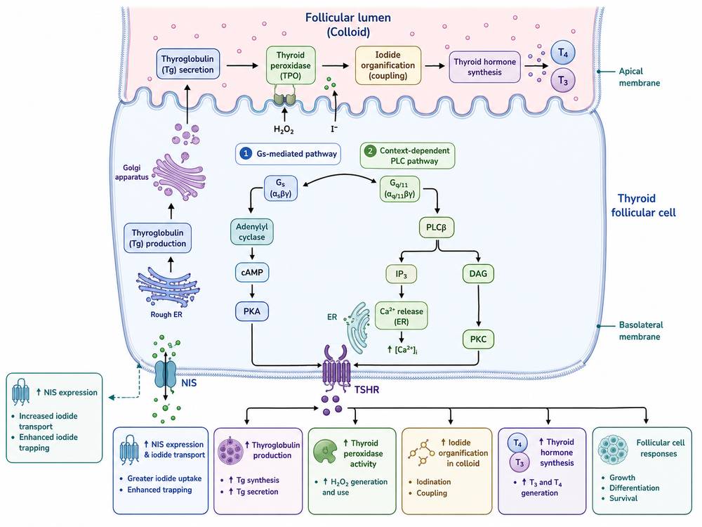
Fig 2. TSHR signaling connects cAMP- and context-dependent phospholipase C pathways with iodide transport, thyroglobulin production, hormone synthesis, and follicular-cell responses.
6. Regulation of Iodide Uptake and Thyroglobulin Production
TSH-stimulated thyroid function coordinates multiple steps of thyroid hormone biosynthesis rather than regulating a single enzyme. At the basolateral membrane, the sodium/iodide symporter (NIS; SLC5A5) concentrates iodide inside the follicular cell. Iodide is then transported toward the apical surface and follicular lumen, where thyroid peroxidase uses hydrogen peroxide generated by the DUOX system to oxidize iodide and attach iodine to selected tyrosine residues in thyroglobulin. Coupling reactions within iodinated thyroglobulin generate T4 and T3 precursors.
Thyroglobulin is synthesized by follicular cells, processed through the endoplasmic reticulum and Golgi, and secreted apically into the follicular lumen. After iodination and storage in colloid, thyroglobulin is endocytosed and proteolyzed, releasing T4 and T3 for secretion across the basolateral membrane. Iodotyrosines that are not coupled into hormone can be deiodinated, allowing iodide recycling.
In vitro, TSH-dependent responses may be measured through NIS or thyroglobulin gene expression, NIS membrane localization, radioiodide or nonradioactive iodide uptake, thyroglobulin secretion, protein iodination, T4/T3 production, and follicle morphology. FRTL-5 studies have shown that thyroglobulin synthesis and iodination can be TSH dependent, but the response is strongly influenced by culture conditions and iodide availability.
| TSH-Regulated Process | Representative Component | Common Experimental Readout | Interpretive Caution |
|---|---|---|---|
| Basolateral iodide uptake | NIS / SLC5A5 | Iodide uptake, NIS mRNA, NIS protein localization | Expression does not guarantee correct membrane trafficking or transport activity |
| Apical iodide handling | Pendrin and related transport mechanisms | Polarized transport or follicle-based assays | Two-dimensional monolayers may not reproduce apical-basolateral polarity |
| Oxidation and organification | TPO, DUOX2/DUOXA2 | Protein-bound iodine, iodinated thyroglobulin, peroxide-dependent activity | Iodide concentration and oxidative stress can alter results |
| Hormone precursor production | Thyroglobulin | TG mRNA, intracellular TG, secreted TG | Secreted quantity and successful iodination are separate endpoints |
| Hormone release | Endocytosis and lysosomal proteolysis | T4/T3 in medium or serum | Requires a sufficiently differentiated and iodide-competent system |
7. T3 and T4 Negative Feedback
Circulating T4 and T3 suppress the HPT axis at both the pituitary and hypothalamic levels. Although T4 is the major thyroidal secretory product, local conversion of T4 to T3 by type 2 deiodinase contributes substantially to feedback signaling in the brain and pituitary. T3 binds nuclear thyroid hormone receptors, particularly thyroid hormone receptor beta isoforms in relevant feedback tissues, and reduces transcriptional and secretory drive for TRH and TSH.
Negative feedback is nonlinear and individualized. Small changes in free T4 can be associated with larger changes in TSH, but the exact relationship depends on the individual set point, age, physiological state, illness, medication exposure, and assay platform. In experimental animals, species-specific hormone-binding proteins, deiodinase patterns, activity cycles, and metabolic rates can further affect the relationship between measured thyroid hormone and TSH.
Feedback experiments should specify whether the endpoint reflects direct pituitary action, hypothalamic regulation, peripheral conversion, or integrated whole-axis behavior. Exogenous T3 may produce a different temporal and tissue pattern from T4 because it bypasses the need for local activation, while T4-based feedback depends in part on tissue-specific uptake and deiodination.
8. Physiological Versus Experimental TSH Responses
Physiological TSH exposure is pulsatile, circadian, and embedded in a closed feedback loop, whereas experimental TSH stimulation is typically delivered under controlled exposure conditions. Laboratory stimulation is often delivered as a fixed concentration, a single bolus, or continuous exposure in a system with limited or absent feedback. This difference can change receptor occupancy, cAMP kinetics, desensitization, gene expression, growth, and functional differentiation.
| Feature | Physiological TSH Exposure | Typical Experimental Exposure | Potential Consequence |
|---|---|---|---|
| Temporal pattern | Pulsatile with circadian modulation | Single addition or continuous incubation | Different receptor adaptation and downstream kinetics |
| Feedback | TRH, TSH, T4, and T3 form a closed regulatory loop | Often absent in isolated cells | Responses may persist beyond physiological limits |
| Ligand form | Endogenous, glycoform-heterogeneous TSH | Purified pituitary or recombinant preparation | Mass concentration may not equal biological potency |
| Target context | Polarized follicles with vascular and stromal support | Monolayer cells or engineered receptor systems | Loss of polarity or thyroid-specific machinery |
| Co-regulators | Insulin/IGF, iodide, neural, immune, and metabolic inputs | Defined medium with selected supplements | Growth and differentiation may become uncoupled |
For cell-based work using bovine TSH for thyroid research, investigators should verify receptor-species compatibility, define activity in the chosen assay, and report both the nominal dose and the response window. A preparation that is highly active in a recombinant human TSHR cAMP assay may behave differently in rat thyroid cells, primary human thyrocytes, or three-dimensional follicles.
9. Common Research Models and Readouts
No single model reproduces every level of TSH biology research. Model selection should follow the biological question: receptor pharmacology, pituitary secretion, thyroid differentiation, iodide transport, hormone synthesis, follicular architecture, or integrated endocrine feedback.
| Research Model | Best-Suited Questions | Typical Readouts | Important Limitations |
|---|---|---|---|
| Recombinant TSHR-expressing HEK or CHO cells | Ligand potency, receptor pharmacology, stimulating or blocking activity | cAMP, reporter gene, calcium, receptor internalization | Limited thyroid-specific differentiation and artificial receptor density |
| FRTL-5 or PCCL3 rat thyroid cells | TSH-dependent differentiation, iodide uptake, NIS/TG regulation, proliferation | cAMP, iodide uptake, NIS, TG, TPO, DNA synthesis | Species-specific response and strong dependence on medium composition |
| Primary human thyrocytes | Human thyroid signaling and differentiated functions | cAMP, gene expression, protein secretion, iodide handling | Donor variability, limited lifespan, variable dedifferentiation |
| Three-dimensional follicles or thyroid organoids | Polarity, lumen formation, organification, hormone biosynthesis | Follicle morphology, apical localization, iodination, T4/T3 | Maturation, standardization, and throughput remain variable |
| Pituitary thyrotroph models | TRH-stimulated secretion and thyroid hormone feedback | TSH release, TSHB expression, calcium, promoter activity | Some lines incompletely reproduce mature thyrotroph physiology |
| Rodent or other in vivo models | Integrated HPT-axis feedback, thyroid growth, endocrine pharmacology | Serum TSH/T4/T3, thyroid weight, histology, gene expression, iodide uptake | Species, sex, age, diet, activity cycle, and iodine intake affect translation |
Readout Timing Matters
- Minutes: cAMP accumulation, calcium changes, receptor phosphorylation, and early kinase activation.
- Hours: immediate-early genes, transcription-factor activity, receptor desensitization, and early NIS/TG transcriptional changes.
- One to several days: iodide uptake capacity, differentiated protein expression, thyroglobulin secretion, proliferation, follicular organization, and hormone synthesis.
Continuous stimulation should not automatically be used for every endpoint. Pulse-chase designs, washout periods, and time-course sampling can reveal transient signaling, delayed differentiation, or receptor adaptation that would be hidden by a single terminal measurement.
10. Key Considerations When Interpreting TSH Biology
TSH biology is sensitive to experimental context. The most reliable studies define the ligand, receptor system, exposure pattern, culture conditions, and endpoint before comparing potency or mechanism.
- Confirm hormone identity and source. Record species, pituitary-derived versus recombinant origin, purity, formulation, glycosylation information when available, and the stated biological activity.
- Do not equate mass with potency. Establish a dose-response curve in the exact assay and report the response metric, such as EC50, fold induction, or activity units.
- Match ligand and receptor species. Cross-species agonism can be useful, but relative potency and receptor coupling may differ across human, bovine, rat, and other systems.
- Control iodide and medium composition. Iodide concentration, serum, insulin/IGF, hydrocortisone, transferrin, and other supplements can alter differentiation, proliferation, and transport.
- Separate receptor activation from thyroid function. A cAMP response confirms proximal signaling but does not prove NIS trafficking, iodide organification, thyroglobulin iodination, or T4/T3 production.
- Account for cell polarity. Monolayer cultures may express thyroid markers without recreating basolateral uptake, apical transport, and follicular lumen chemistry.
- Use multiple time points. Early signaling and delayed biosynthetic responses occur on different timescales and may show different dose dependencies.
- Monitor desensitization and passage effects. Receptor density, chronic TSH exposure, cell passage, and dedifferentiation can reduce or reshape responses.
- Include orthogonal controls. Forskolin can test adenylyl cyclase competence; a TSHR antagonist or receptor-negative cell can support specificity; vehicle and positive controls help detect assay drift.
- Interpret endocrine measurements with assay limitations in mind. TSH and thyroid hormone immunoassays can be affected by platform differences, binding proteins, heterophile antibodies, macro-TSH, and other analytical interference.
TSH is best understood as a context-dependent endocrine signal that links central feedback control to thyroid-cell differentiation and hormone biosynthesis. Mechanistic conclusions are strongest when proximal receptor signaling is paired with functional thyroid readouts and when the experimental hormone preparation is qualified in the same biological system in which it will be used.
References
- Fekete C, Lechan RM. Central regulation of hypothalamic-pituitary-thyroid axis under physiological and pathophysiological conditions. Endocr Rev. 2014;35(2):159-194. doi:10.1210/er.2013-1087.
- Ortiga-Carvalho TM, Chiamolera MI, Pazos-Moura CC, Wondisford FE. Hypothalamus-pituitary-thyroid axis. Compr Physiol. 2016;6(3):1387-1428. doi:10.1002/cphy.c150027.
- Szkudlinski MW, Fremont V, Ronin C, Weintraub BD. Thyroid-stimulating hormone and thyroid-stimulating hormone receptor structure-function relationships. Physiol Rev. 2002;82(2):473-502. doi:10.1152/physrev.00031.2001.
- Rapoport B, Chazenbalk GD, Jaume JC, McLachlan SM. The thyrotropin (TSH) receptor: interaction with TSH and autoantibodies. Endocr Rev. 1998;19(6):673-716. doi:10.1210/edrv.19.6.0352.
- Nilsson M. Iodide handling by the thyroid epithelial cell. Exp Clin Endocrinol Diabetes. 2001;109(1):13-17. doi:10.1055/s-2001-11014.
- Bizhanova A, Kopp P. Minireview: the sodium-iodide symporter NIS and pendrin in iodide homeostasis of the thyroid. Endocrinology. 2009;150(3):1084-1090. doi:10.1210/en.2008-1437.
- Ikegami K, Refetoff S, Van Cauter E, Yoshimura T. Interconnection between circadian clocks and thyroid function. Nat Rev Endocrinol. 2019;15(10):590-600. doi:10.1038/s41574-019-0237-z.
- Kimura T, Van Keymeulen A, Golstein J, Fusco A, Dumont JE, Roger PP. Regulation of thyroid cell proliferation by TSH and other factors: a critical evaluation of in vitro models. Endocr Rev. 2001;22(5):631-656. doi:10.1210/edrv.22.5.0444.
- Ossendorp FA, Leer LM, Bruning PF, van den Brink JAM, Sterk A, de Vijlder JJM. Iodination of newly synthesized thyroglobulin by FRTL-5 cells is selective and thyrotropin dependent. Mol Cell Endocrinol. 1989;66(2):199-205. doi:10.1016/0303-7207(89)90032-4.
- Van Uytfanghe K, Ehrenkranz J, Halsall D, et al. Thyroid stimulating hormone and thyroid hormones (triiodothyronine and thyroxine): an American Thyroid Association-commissioned review of current clinical and laboratory status. Thyroid. 2023;33(9):1013-1028. doi:10.1089/thy.2023.0169.
- Kang HS, Kumar D, Liao G, et al. GLIS3 is indispensable for TSH/TSHR-dependent thyroid hormone biosynthesis and follicular cell proliferation. J Clin Invest. 2017;127(12):4326-4337. doi:10.1172/JCI94417.